Posts tagged #emergency medicine

Stingray Stings

Written by: Mike Tandlich, MD (NUEM ‘24) Edited by: Peter Serina, MD, MPH (NUEM ‘22)
Expert Commentary by: Mike Macias, MD (NUEM ‘17)


Expert Commentary

Thank you Drs. Tandlich and Serina for this excellent infographic summarizing stingray envenomation! The good news is that the majority of stingray injuries are nonfatal and will heal without any complications! You hit all of the key points however I just wanted to highlight a few management tips below: 

Treat as a Trauma! 

While majority of the pain from stingray envenomation occurs as a result of its venom, it is important to remember that this is also a traumatic injury. Treat the injury just like you would any other penetrating trauma. Consider the location as well as surrounding structures and make sure to properly examine for tendon, nerve, and vascular injury. Injuries to the chest or abdominal regions should prompt advanced imaging and trauma consultation. 

Hot Water is Key! 

Stingray envenomation is noted to cause severe pain that is often out of proportion to your examination findings. While the exact mechanism is not clear, the venom can lead to not only pain but also local tissue necrosis. The good news is the venom is heat labile! The faster you can get the injured area into hot water the better. You want the water to be as hot as tolerable without causing a thermal burn. A good rule of thumb is to have the patient place their unaffected limb in the water first to see if it is tolerable. As this often occurs at a beach, lifeguards are often your best resource to get hot water fast! Oral analgesics can be administered if needed however often they are unnecessary as soon as the injured area is submerged in hot water. 

Retained Barb?

While uncommon, a retained barb from the envenomation can occur so be sure to consider this and evaluate appropriately. Traditionally, x-ray imaging of the affected area is performed to evaluate for a radio-opaque barb however some evidence suggests this to be a relatively low yield practice [1]. Ultrasound can also be considered if there is suspicion for retained barb or other material. In general ultrasound has been shown to be highly sensitive for identification of foreign body [2]. Not only can it be used to identify the barb but it can be used to facilitate removal [3]. 

Give Prophylactic Antibiotics 

Prophylactic antibiotics are recommended for stingray envenomation given that the limited data suggest a higher rate of wound infection in patients who were not initially treated with antibiotics [1]. Given these injuries often occur in the ocean make sure to cover for salt water species such as Vibro. Levofloxacin is my go to option.

Teach The Stingray Shuffle! 

Keeping these key management points in mind, the good news is that the majority of stingray injuries are nonfatal and will heal without any complications! Before your patient is discharged don’t forget to remind them that the next time they are going out for a surf to do the stingray shuffle!

References

  1. Clark RF, Girard RH, Rao D, Ly BT, Davis DP. Stingray envenomation: a retrospective review of clinical presentation and treatment in 119 cases. J Emerg Med. 2007 Jul;33(1):33-7

  2. Aras MH, Miloglu O, Barutcugil C, Kantarci M, Ozcan E, Harorli A. Comparison of the sensitivity for detecting foreign bodies among conventional plain radiography, computed tomography and ultrasonography. Dentomaxillofac Radiol. 2010;39(2):72-78. doi:10.1259/dmfr/68589458

  3. Nwawka OK, Kabutey NK, Locke CM, Castro-Aragon I, Kim D. Ultrasound-guided needle localization to aid foreign body removal in pediatric patients. J Foot Ankle Surg. 2014;53(1):67-70. doi:10.1053/j.jfas.2013.09.006

Michael Macias, MD

Systems Clinical Ultrasound Director,
Emergent Medical Associates

Ultrasound Director,
UHS SoCal MEC Residency Programs


How To Cite This Post:

[Peer-Reviewed, Web Publication] Tandlich, M. Serina, P. (2021, Nov 15). Stingray Stings. [NUEM Blog. Expert Commentary by Macias, M]. Retrieved from http://www.nuemblog.com/blog/stingray-stings


Other Posts You May Enjoy

Posted on November 15, 2021 and filed under Environmental.

Buprenorphine Use in the ED

Written by: Diana Halloran, MD (NUEM ‘24) Edited by: Sean Watts, MD (NUEM ‘22) Expert Commentary by: Quentin Reuter, MD (NUEM ‘18)

Written by: Diana Halloran, MD (NUEM ‘24) Edited by: Sean Watts, MD (NUEM ‘22) Expert Commentary by: Quentin Reuter, MD (NUEM ‘18)


The United States has been facing a debilitating opioid epidemic, which has been partially fueled by the over-prescription of these medications in the emergency department setting. In addition, the opioid epidemic has grown exponentially during the COVID-19 pandemic. More than 40 states have reported increases in opioid-related mortality, resulting in an increased burden on an already overstrained healthcare system. (1) Prescribing the medication Buprenorphine in the emergency department offers an opportunity to ameliorate these past faults and rising statistics.

The basics:

Buprenorphine, which goes by the trade name Subutex, works by acting as both a partial mu agonist and weak kappa antagonist on opiate receptors in the brain. (2) This mechanism of action enables buprenorphine to exert analgesic effects, as well as antagonistic effects when additional opiates are consumed. In addition, buprenorphine does not carry significant sedative effects, making respiratory depression extremely rare. (3) Buprenorphine is also safe in pregnancy – a 2016 meta-analysis found no difference in pregnant patients given methadone versus buprenorphine when assessing for congenital malformations. (4) The American College of Obstetrics & Gynecology has released a committee position statement, encouraging the use of buprenorphine in pregnant patients with opioid use disorder. (5)

How to prescribe:

While the DEA X-waiver is required to write a prescription for buprenorphine for addiction treatment, withdrawal, or detox, it is not required to order or administer a dose in the hospital or emergency department. (6) This exception, called the “three-day rule”, allows a patient to come to the emergency department for three consecutive days to obtain a dose of buprenorphine if found to be in opioid withdrawal. (7)

In order to dose buprenorphine in the emergency department, the patient must be in mild acute opioid withdrawal, with a Clinical Opiate Withdrawal Score (COWS) of at least 8. (8,9) Administration of buprenorphine should not occur if the patient does not appear to be clinically withdrawing, as administration in this setting could actually precipitate withdrawal.

Dosing: (10)

  • 4mg of sublingual buprenorphine can be given initially, allowing 20-40 minutes for resolution of withdrawal symptoms with repeat dosing every 1-2 hours as needed. (10)

  • On Day 2, the patient’s response to Day 1 should be assessed. If the patient’s opioid withdrawal symptoms were controlled, the same dose can be continued. If not, the dose should be increased by 2-4mg. (10)

  • On Day 3, the patient’s response to Day 2 should be assessed. Again, if the patient’s withdrawal symptoms are controlled then the same dose can be continued. If not, the dose can be increased by 2-4mg for Day 3. (10)

  • After 3 days this dose should be continued for 3-7 days until steady-state levels are achieved (10)

  • Doses should be decreased by 2mg if the patient experiences opioid intoxication (10)

Use in the emergency department:

While buprenorphine and long-term treatment of opioid use disorder may seem confined to primary care physicians and psychiatrists, emergency medicine physicians have been shown to be successful providers for initiating buprenorphine treatment versus brief intervention and referral with a result of decreased self-reported illicit opioid use. (11) In addition, Dr. Gail D’Onofrio, chair of the Department of Emergency Medicine at Yale, found that emergency department initiated buprenorphine treatment was associated with the increased self-reported engagement of addiction treatment and reduced illicit opioid use within a two-month interval. (12)  Increasing evidence demonstrates that the emergency department provides an opportunity to intervene on opioid use disorder, with more and more emergency medicine physicians becoming X-waiver certified.

References

  1. Issue brief: Reports of increases in opioid and other drug-related overdose and other concerns during COVID pandemic. American Medical Association. https://www.ama-assn.org/system/files/2020-12/issue-brief-increases-in-opioid-related-overdose.pdf. Published December 9, 2020.

  2. Wakhlu S. Buprenorphine: a review. J Opioid Manag. 2009 Jan-Feb;5(1):59-64. doi: 10.5055/jom.2009.0007.

  3. Walsh SL, Preston KL, Stitzer ML, Cone EJ, Bigelow GE. Clinical pharmacology of buprenorphine: ceiling effects at high doses. Clin Pharmacol Ther. 1994 May;55(5):569-80. doi: 10.1038/clpt.1994.71.

  4. Zedler BK, Mann AL, Kim MM, Amick HR, Joyce AR, Murrelle EL, Jones HE. Buprenorphine compared with methadone to treat pregnant women with opioid use disorder: a systematic review and meta-analysis of safety in the mother, fetus and child. Addiction. 2016 Dec;111(12):2115-2128. doi: 10.1111/add.13462.

  5. Committee Opinion No. 711 Summary: Opioid Use and Opioid Use Disorder in Pregnancy. Obstetrics & Gynecology. 2017;130(2):488-489. doi:10.1097/aog.0000000000002229

  6. Special Circumstances for Providing Buprenorphine. SAMHSA. https://www.samhsa.gov/medication-assisted-treatment/statutes-regulations-guidelines/special-circumstances. Published August 19, 2020.

  7. Nagel L. Emergency Narcotic Addiction Treatment. https://www.deadiversion.usdoj.gov/pubs/advisories/emerg_treat.htm.

  8. Wesson DR, Ling W. Clinical Opiate Withdrawal Scale. PsycTESTS Dataset. June 2003. doi:10.1037/t48752-000

  9. D'Onofrio G, O'Connor PG, Pantalon MV, et al. Emergency department-initiated buprenorphine/naloxone treatment for opioid dependence: a randomized clinical trial. JAMA. 2015;313(16):1636-1644. doi:10.1001/jama.2015.3474

  10. Dosing Guide For Optimal Management of Opioid Dependence. The National Alliance of Advocates for Buprenorphine Treatment.

  11. D’Onofrio G, O’Connor PG, Pantalon MV, et al. Emergency Department–Initiated Buprenorphine/Naloxone Treatment for Opioid Dependence: A Randomized Clinical Trial. JAMA. 2015;313(16):1636–1644. doi:10.1001/jama.2015.3474

  12. D'Onofrio G, Chawarski MC, O'Connor PG, Pantalon MV, Busch SH, Owens PH, Hawk K, Bernstein SL, Fiellin DA. Emergency Department-Initiated Buprenorphine for Opioid Dependence with Continuation in Primary Care: Outcomes During and After Intervention. J Gen Intern Med. 2017 Jun;32(6):660-666. doi: 10.1007/s11606-017-3993-2.


Expert Commentary

Thanks to Dr. Halloran and Watts for providing an informative discussion on buprenorphine prescribing from the ED. Buprenorphine continues to emerge as the state of the art treatment strategy for opioid use disorder (OUD) and thus, developing a working knowledge for when and how to use it is essential.

While there is little doubt that the medical field fueled the opioid epidemic through the prescribing of pain medications, EM is often given a disproportionate amount of blame for the current situation.  In 2012, EM prescriptions made up only 4.3% of all opioids in circulation (1). Furthermore, I anticipate our specialty will continue to lead the fight against the opioid epidemic as practices such as naloxone prescribing, education around safe injecting practices, reduction and optimization of opioid prescribing efforts, and buprenorphine initiation gain further traction in the ED.

Obtaining a DEA X is the first step to prescribing buprenorphine. In April of this year guidelines for the administration of buprenorphine were updated to allow practitioners to treat up to 30 patients at a time with no extra training (2). While these changes will likely expand buprenorphine prescribing from the ED, it is vital that we do not operate in a silo.

To effectively manage this complex patient cohort, a coherent system of addiction medicine services is vital.  EDs must partner with local community resources to make rapid addiction medicine appointments available. Our department utilizes specially trained addiction care coordinators, nurses with extensive training in addiction medicine to help evaluate OUD patients and navigate the fractured array of outpatient services.

Prior to the implementation of our Medication for Opioid Use Disorder (MOUD) program, our clinicians had relatively little to offer patients that directly addressed their underlying addiction.  While anecdotal, we believe that by utilizing MOUD, we have begun to rebuild trust between OUD patients and the medical system.  A once generally negative relationship between OUD patients and our ED staff has been replaced with a hopeful rapport, confident that recovery for these patients is a distinct possibility.  This therapeutic relationship continues to grow and we believe will lead to long-term sustained recovery for many of our OUD patients in the surrounding community. 

References

  1. Levy B, Paulozzi L, Mack KA, Jones CM. Trends in Opioid Analgesic-Prescribing Rates by Specialty, U.S., 2007-2012. Am J Prev Med 2015;49:409-13.

  2. Reuter Q, Smith G, McKinnon J, Varley J, Jouriles N, Seaberg D. Successful Medication for Opioid Use Disorder (MOUD) Program at a Community Hospital Emergency Department. Acad Emerg Med 2020.

quentin reuter.png

Quentin Reuter, MD

Emergency Medicine Physician

Core Faculty at Summa Health


How To Cite This Post:

[Peer-Reviewed, Web Publication] Halloran D., Watts S. (2021, Sept 13). Buprenorphine Use in the ED. [NUEM Blog. Expert Commentary by Reuter Q.]. Retrieved from http://www.nuemblog.com/blog/buprenorphine


Other Posts You May Enjoy

Basic Capnography Interpretation

Written by: Shawn Luo, MD (NUEM ‘22) Edited by: Matt McCauley, MD (NUEM ‘21) Expert Commentary by: N. Seth Trueger, MD, MPH

Written by: Shawn Luo, MD (NUEM ‘22) Edited by: Matt McCauley, MD (NUEM ‘21) Expert Commentary by: N. Seth Trueger, MD, MPH


Continuous waveform capnography has increasingly become the gold standard of ETT placement confirmation. However, capnography can provide additional valuable information, especially when managing critically ill or mechanically ventilated patients.

Normal Capnography

  • Phase I (inspiratory baseline) reflects inspired air, which is normally devoid of CO2.

  • Phase II (expiratory upstroke) is the transition between dead space to alveolar gas.

  • Phase III is the alveolar plateau. Traditionally, PCO2 of the last alveolar gas sampled at the airway opening is called the EtCO2. (normally 35-45 mmHg)

  • Phase 0  is the inspiratory downstroke, the beginning of the next inspiration

Figure 1. Normal Capnography Tracing (emDOCs.net)

EtCO2 is only one component of capnography. Measured at the end-peak of each waveform, it reflects alveolar CO2 content and is affected by alveolar ventilation, pulmonary perfusion, and CO2 production.

Figure 2. Factors affecting ETCO2 (EMSWorld)

Figure 2. Factors affecting ETCO2 (EMSWorld)

EtCO2 - PaCO2 Correlation

Correlating EtCO2 and PaCO2 can be problematic, but in general, PaCO2 is almost always HIGHER than EtCO2. Normally the difference should be 2-5mmHg but the PaCO2-EtCO2 gradient is often increased due to increased alveolar dead space (high V/Q ratio), such as low cardiac output, cardiac arrest, pulmonary embolism, high PEEP ventilation.

Important Patterns

Let’s go through a few cases and learn some of the important capnography waveforms to recognize

Case 1: Capnography with Advanced Airway

An elderly gentleman with a history of COPD, CAD & CKD gets rushed into the trauma bay with respiratory distress and altered mental status. You gave him a trial of BiPAP for a few minutes without improvement.

  1. You swiftly tubed the patient. It was not the easiest view, but you advance the ETT hoping for the best. Upon attaching the BVM to bag the patient, you saw this on capnography:

Figure 3. Case 1 (EMSWorld)

Figure 3. Case 1 (EMSWorld)

Oops, the ETT is in the esophagus, as evidenced by the low-level EtCO2 that quickly tapers off.

2. You remove the ETT, bag the patient up, and try again with a bougie. Afterward, you see…

Figure 4. Capnography with ETT in right main bronchus (EMSWorld)

Figure 4. Capnography with ETT in right main bronchus (EMSWorld)

This suggests a problem with ETT position, most often in the right main bronchus. Notice the irregular plateau--the initial right lung ventilation, followed by CO2 escaping from the left lung. Beware that capnography can sometimes still appear normal despite the right main bronchus placement.

3. You pull back the ETT a few cm and the CXR now confirms the tip is now above the carina. The patient’s capnography now looks like this:

Figure 5. Capnography showing obstruction or bronchospasm (SketchyMedicine)

Figure 5. Capnography showing obstruction or bronchospasm (SketchyMedicine)

Almost looks normal but notice the “shark fin” appearance, this is due to delayed exhalation, often seen in airway obstruction and bronchospasms such as COPD or asthma exacerbation.

4. You suction the patient and administer several bronchodilator nebs. The waveform now looks more normal:

Figure 6. Capnography showing normal waveform (SketchyMedicine)

5. However, just as you were about to get back to the workstation to call the ICU, the monitor alarms and you see this:

Figure 7. Sudden loss of capnography waveform (SketchyMedical)

Figure 7. Sudden loss of capnography waveform (SketchyMedical)

Noticing the ETT still in place with good chest rise, you quickly check for a pulse. There is none.

6. You holler, push the code button and start ACLS with a team of clinicians. With CPR in progress, you notice this capnography:

Figure 8. Capnography during CPR (SketchyMedicine)

Figure 8. Capnography during CPR (SketchyMedicine)

Initially, your patient’s EtCO2 was only 7, after coaching the compressor and improving CPR techniques, it increased to 14.

You are also aware that EtCO2 at 20min of CPR has prognostic values. EtCO2 <10 mmHg at 20 minutes suggests little chance of achieving ROSC and can be used as an adjunctive data point in the decision to terminate resuscitation.

7. Fortunate for your patient, during the 3rd round of ACLS, you notice the following:

Figure 9. ROSC on capnography (emDOCs.net)

Figure 9. ROSC on capnography (emDOCs.net)

This sudden jump in EtCO2 suggests ROSC. You stop the CPR and confirm that the patient indeed has a pulse.

8. As you are putting in orders for post-resuscitation care, you notice this:

Figure 10. Asynchronous breathing on capnography (SketchyMedical)

Figure 10. Asynchronous breathing on capnography (SketchyMedical)

This curare cleft comes from the patient inhaling in between ventilator-delivered breaths and is usually a sign of asynchronous breathing. However, in the post-arrest scenario, it is a positive prognostic sign as your patient is breathing spontaneously. You excitedly call your mom, I meant MICU, about the incredible save. 

Case 2: Capnography with Non-intubated Patient

You just hung up the phone with MICU when EMS brings you a young woman with a heroin overdose. She already received some intranasal Narcan from EMS but per EMS report patient is becoming sleepy again.

  1. She mumbles a little as you shout her name, and as you put an end-tidal nasal cannula on her, you saw this:

Figure 11. Hypoventilation on capnography (emDOCs.net)

Figure 11. Hypoventilation on capnography (emDOCs.net)

Noticing the low respiratory rate and high EtCO2 value, you recognize this is hypoventilation.

2. But very soon she becomes even less responsive and the waveform changed again:

Figure 12. Airway obstruction on capnography (emDOCs.net)

Figure 12. Airway obstruction on capnography (emDOCs.net)

The inconsistent, interrupted breaths suggest airway obstruction, while the segments without waveform suggest apnea. You have to act fast.

3. By then your nurse has already secured an IV, so you pushed some Narcan. However, in the heat of the moment, you gave the whole syringe. The patient quickly woke up crying and shaking.

Figure 13. Hyperventilating on capnography (emDOCs.net)

Figure 13. Hyperventilating on capnography (emDOCs.net)

She was quite upset and hyperventilating. The waveform reveals a high respiratory rate and relatively low EtCO2.

As much as you are a little embarrassed by putting the patient into florid withdrawal, you know it could have been a lot worse. Walking away from the shift, you think about how many times capnography has assisted you during those critical moments. “Hey, perhaps we should buy a capnography instead of a baby monitor,” you ask your wife at dinner.

Additional Resources

This website provides a tutorial and quiz on some of the basic capnography waveforms.

References

  1. American Heart Association. 2019 American Heart Association Focused Update on Advanced Cardiovascular Life Support. Circulation. 2019; 140(24). https://doi.org/10.1161/CIR.0000000000000732

  2. Brit Long. Interpreting Waveform Capnography: Pearls and Pitfalls. emDOCs.net. www.emdocs.net/interpreting-waveform-capnography-pearls-and-pitfalls/, accessed May 12, 2020

  3. Capnography.com, accessed May 12, 2020

  4. Kodali BS. Capnography outside the operating rooms. Anesthesiology. 2013 Jan;118(1):192-201. PMID: 23221867.

  5. Long, Koyfman & Vivirito. Capnography in the Emergency Department: A Review of Uses, Waveforms, and Limitations. Clinical Reviews in Emergency Medicine. 2017; 53(6). https://doi.org/10.1016/j.jemermed.2017.08.026

  6. Nassar & Schmidt, Capnography During Critical Illness. CHEST. 2016; 249(2). https://doi.org/10.1378/chest.15-1369

  7. Sketchymedicine.com/2016/08/waveform-capnography, accessed May 13, 2020

  8. Wampler, D. A. Capnography as a Clinical Tool. EMS World. www.emsworld.com/article/10287447/capnography-clinical-tool. June 28, 2011. Accessed May 13, 2020


Expert Commentary

This is a nice review of many of the intermediate and qualitative uses of ETCO2 in the ED. For novices, I recommend a few basic places to start:

  1. Confirmation of intubation. Color change is good but it’s just litmus paper and gets easily defeated by vomit. Also, in low output states, it may not pick up. Further, colorimetric capnographs require persistent change over 6 breaths, not just a single change. Waveform capnography uses mass spec or IR spec to detect CO2 molecules. There are so many uses, it’s good to have, I don’t see why some are resistant to use this better plastic adapter connected to the monitor vs the other, worse, plastic adapter.

a. The mistake I have seen here is assuming a lack of waveform is due to low cardiac output, ie there’s no waveform because the patient is being coded, not because of esophageal intubation. There is always *some* CO2 coming out if there is effective CPR; if there isn’t, the tube is in the wrong place. If you really don’t believe it, check with good VL but a flatline = esophagus.

2. Procedural sedation. There’s lots of good work and some debate about absolute or relative CO2 changes or qualitative waveform changes that might predict impending apnea, but for me, the best use is that I can just glance at the monitor for a second or two and see yes, the patient is breathing. No more staring at the chest debating whether I see chest rise, etc. It’s like supervising a junior trainee during laryngoscopy with VL: it’s anxiolysis for me.

a. Using ketamine? Chest movement or other signs of respiratory effort without ETCO2 waveform means laryngospasm. Jaw thrust, bag, succinylcholine (stop when better).

3. Cardiac arrest.

a. Quality of CPR. Higher number means more output. Can mean the compressor needs to fix their technique, or more often, is tiring out and needs a swap.

b. ROSC. There can be a big jump (eg from 15 to 40) when ROSC occurs. Very helpful.

c. Ending a code. 20 mins into a code, if it’s <10 during good CPR, the patient is unlikely to survive. I try to view this as confirming what we know – it’s time to end the code. The mistake here is to not end a code that should otherwise end because the ETCO2 is above 10; it doesn’t work like that, it’s a 1-way test.

4. Leak. One waveform shape I wanted to add that I find helpful: if the downstroke kinda dribbles down like a messy staircase, it’s a leak. Can be an incomplete connection (eg tubing to the vent) or the balloon is too empty or full.

Seth Trueger, MD, MPH

Assistant Professor of Emergency Medicine

Department of Emergency Medicine

Northwestern University


How To Cite This Post:

[Peer-Reviewed, Web Publication] Luo, S., McCauley M. (2021, Sept 9). Basic Capnography Interpretation. [NUEM Blog. Expert Commentary by Trueger N.S]. Retrieved from http://www.nuemblog.com/blog/capnography


Other Posts You May Enjoy

Droperidol

Written by: Adam Payne, MD (NUEM ‘24) Edited by: Julian Richardson, MD (NUEM ‘21) Expert Commentary by: Matt O' Connor, MD

Written by: Adam Payne, MD (NUEM ‘24) Edited by: Julian Richardson, MD (NUEM ‘21) Expert Commentary by: Matt O' Connor, MD


Expert Commentary

Thanks to Dr. Payne & Dr. Richardson for putting this together!  I think this was well done, they’ve presented a concise overview of the safety and efficacy of droperidol. 

There’s a lot of utility in droperidol.  It’s great for nausea, migraines, and even as an adjunct for chronic pain.  It’s also a very good choice for agitation.  I use it most often for nausea.  It’s been shown to be as effective as odansetron, and more effective than metoclopramide.  Anecdotally, I find it works particularly well for gastroparesis and cannabinoid hyperemesis (with some low-concentration topical capsaicin cream), with less sedation than haloperidol.  For migraines, it has been shown to be as effective as prochlorperazine.  It works well for sedation in agitated patients as well; IV & IM it has a much faster onset than haloperidol, and so benzodiazepines typically do not have to be co-administered, reducing the level and duration of sedation and need for monitoring.     

The black box warning significantly limited droperidol’s availability, such that many of our newer graduates have not had any first-hand clinical experience with the medication.  If you’re not familiar with its use, don’t let the black box warning completely dissuade you.  Subsequent studies looking at emergency department droperidol use have shown it to be safe, and that complications related to QT prolongation are rare in typical doses.   As a rule of thumb, the dose of droperidol is about half of the dose of haloperidol for a given indication.  For nausea, migraine, or other pain, I usually start with 0.625-2.5mg IV, twice that IM, and can repeat dosing if needed (my most common starting dose is 1.25mg IV).  For agitation, usually 2.5-5mg IM, though up to 10mg IM has been shown likely to be safe.  Although it is prudent to be cautious, I think the literature supports droperidol’s use at appropriate doses in otherwise healthy patients.

matt oconnor.PNG

Matt O’Connor, MD

Emergency Medicine Physician

BerbeeWalsh Department of Emergency Medicine

University of Wisconsin Hospitals and Clinics


How To Cite This Post:

[Peer-Reviewed, Web Publication] Payne, A. Richardson, J. (2021, Aug 30). Droperidol. [NUEM Blog. Expert Commentary by O’Connor, M]. Retrieved from http://www.nuemblog.com/blog/droperidol


Other Posts You May Enjoy

Awake Intubation

Written by: Patricia Bigach MD, (NUEM ‘23) Edited by: Terese Whipple (NUEM ‘20) Expert Commentary by: Seth Trueger MD, MPH

Written by: Patricia Bigach MD, (NUEM ‘23) Edited by: Terese Whipple (NUEM ‘20) Expert Commentary by: Seth Trueger MD, MPH


Awake Intubation Final.png

Expert Commentary

Awake intubation can sound imposing but simply means the patient is still breathing on their own. It is mostly just a matter of using topical lidocaine instead of paralyzing, and sedating the patient a bit to tolerate it. It will almost always require some sedation – ketamine procedural sedation works very well as the patient’s protective reflexes will be intact (until we topicalize) as will their respiratory drive.

It does not take long! Just spray lido instead of pushing NMBA. This is the key concept. If time is really a factor, I atomize the larynx, push ketamine, and then reload and spray more lidocaine as I do laryngoscopy; everything else is just like every other ED intubation.

Glycopyrrolate is nice but if it’s not handy, not worth a delay.

I find nebulizing doesn’t add much, mostly just gets the mouth. I still nebulize if I can get it set up quickly while prepping everything else (and it can help tolerate the atomizer).

Small touches of propofol might help relax the ketamine-sedated patient as well, including spontaneous/dissociated movements and tightly closed mouths. Dexmedetomidine might not be fast enough for ED intubations.

I usually use hyperangulated VL (eg Glidescope S3) – we are usually doing this for predicted difficult intubation, and now not optimizing intubating conditions. Fiberoptic requires a fair amount of skill and time. One of the main things that demystified awake intubation for me is it is a medication choice; it doesn’t always mean awake-fiber optic.

In non-COVID times, I would keep the nasal cannula on at 5-15lpm to keep the patient as oxygenated as possible, which is even better than during RSI because they’re still breathing, now with extra oxygen.

The paradox of awake intubation is that we take the patients we predict to be the most difficult anatomically, and then don’t optimize intubating conditions (no NMBA). Part of the beauty of awake intubation is that we also gain a ton of information even if unsuccessful without losing much; if I get a partial view in non-NMBA circumstances I can make a judgment call about proceeding to paralysis (ie RSI) or calling for help, etc.

Sedation-only or ketamine-only intubation can sound like a good idea but neither makes sense to me. It takes a lot of sedation to knock out protective airway reflexes to allow laryngoscopy, i.e. enough to impair respiratory drive. Topicalization is not hard with atomizers. Similarly, ketamine keeps the airway reflexes intact, which is why it is so safe for procedural sedation, so hard to imagine that laryngoscopy won’t be an issue.

Seth Trueger, MD, MPH

Assistant Professor of Emergency Medicine

Department of Emergency Medicine

Northwestern University


How To Cite This Post:

[Peer-Reviewed, Web Publication] Bigach, P. Whipple, T. (2021, Aug 20). Awake Intubation. [NUEM Blog. Expert Commentary by Trueger, S]. Retrieved from http://www.nuemblog.com/blog/awake-intubation


Other Posts You May Enjoy

Posted on August 23, 2021 and filed under Airway.

TPA in Frostbite

Written by: Patrick King, MD Edited by: Nery Porras, MD (NUEM ‘21) Expert Commentary by: Anne Lambert Wagner, MD


TPA in Frostbite

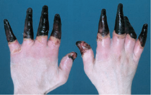
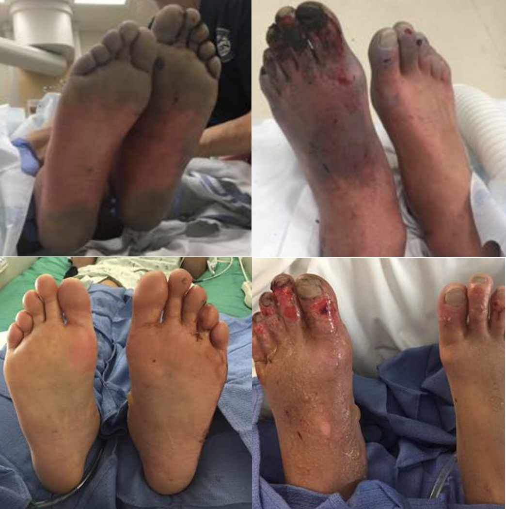
Figure 1. What we would like to avoid (Cline et al.)

Figure 1. What we would like to avoid (Cline et al.)

It’s an early Saturday morning, and EMS brings in one of your ED’s regulars – a schizophrenic, undomiciled gentleman named Jack who finds occasional work as a day laborer. You walk to bed three to greet Jack who is uncomfortable and shivering while nursing collects vitals. His chief complaint is hand and foot pain. You listen to him speak, but you jump right into a cursory exam as he does – and your heart sinks when you see the icy hard, cyanotic, mottled digits across all four extremities. You wonder what else you might be able to offer in addition to the standard cold injury approach we are taught as emergency residents, and you recall that the What’s New in Emergency Medicine section of UpToDate just recognized growing evidence for yet another off-label use for tPA: severe frostbite.

As we head into the winter months, emergency physicians will continue to see frostbite wreck a significant level of morbidity on our most vulnerable patients – patients who are undomiciled, suffering from addictions or mental illness, and those with preexisting conditions that limit blood flow to extremities (Zafren and Crawford Mechem). This post will address the theory, evidence, and logistics behind tPA utilization in severe frostbite.

The proposed efficacy of tPA in frostbite is related to cold-induced thrombosis. Endothelial damage is sustained both as a direct result of cold-related injury and exacerbated by reperfusion injury during the period of rewarming. During rewarming, arachidonic acid cascades promote vasoconstriction, platelet aggregation, leukocyte sludging, and erythrostasis which further promote thrombosis throughout affected tissues. This process is compounded in instances of multiple freeze-thaw cycles (Cline et al).

Research on tPA in frostbite goes back years. In 2005, Twomey et al. demonstrated in an open-label study that technetium (Tc)-99m scintigraphy (i.e., nuclear bone scan) reliably predicts digits/limbs at risk for amputation. Historical control patients with no or minimal flow distal to radiographically identified “cutoff” points of ischemia on bone scans inevitably all required amputations. Untreated historical controls without flow cutoffs were more likely to retain digits. In contrast, 16 of 19 study patients with identified flow cutoffs responded to intra-arterial (IA) or intravenous (IV) tPA with an amputation rate of only 19% of at-risk digits. In 2017, Patel et al. showed a 15% amputation rate for severe frostbite in eight IA tPA patients compared to 77% in their control group.

Figure 2. Pre-tPA and Post-tPA using technetium (Tc)-99m scintigraphy bone scan (Twomey et al.)

Figure 2. Pre-tPA and Post-tPA using technetium (Tc)-99m scintigraphy bone scan (Twomey et al.)


While study results have been impressive in instances of small sample sizes such as the above, a paucity of evidence has prevented widespread utilization of tPA for frostbite use amongst emergency physicians. This year, however, What’s New In Emergency Medicine on UpToDate gave special attention to a 2020 systematic review of 16 studies by Lee and Higgins which wielded a sample size of 209 patients with 1109 digits at high amputation risk. The study, entitled “What Interventional Radiologists Need to Know About Managing Severe Frostbite”, ultimately demonstrated a 76% salvage rate amongst IA tPA (222 amputations amongst 926 digits) and 62% salvage rate in IV tPA (24 amputations amongst 63 patients). Importantly, the 16 studies are not randomized, though several such as Patel et al. and Twomey et al. utilize historical controls. There is also no direct comparison of IA vs. IV tPA, and for unclear reasons, the salvage rate for IA is in terms of digits salvaged out of those at risk while IV is expressed as a function of patients who required no amputations. Though there remains additional research to be done, UpToDate’s Frostbite authors Zafren and Crawford Mechem now give an overall grade 2C recommendation for tPA use in severe frostbite for patients otherwise at risk of life-altering amputations.

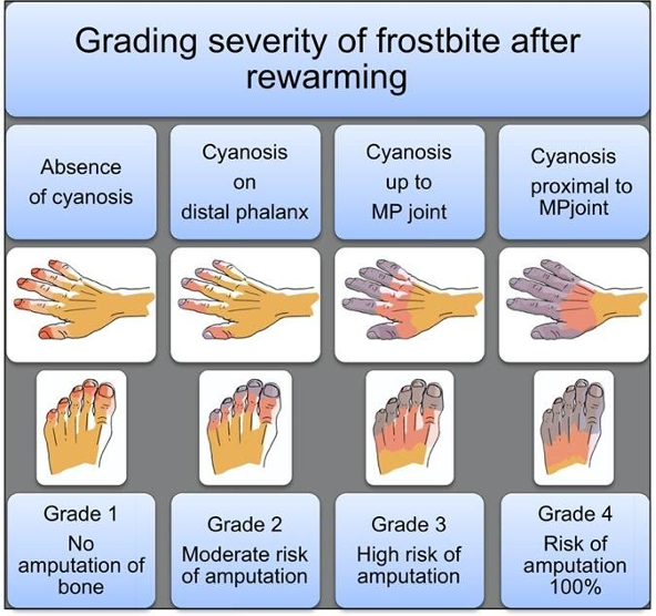
Figure 3. Grading severity of frostbite after rewarming (Cauchy et al.)

Figure 3. Grading severity of frostbite after rewarming (Cauchy et al.)

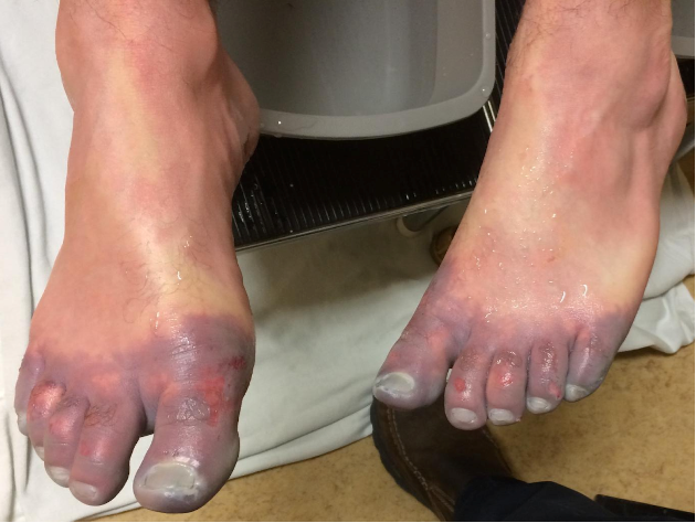
Figure 4. Grade 4 Frostbite, best seen in far right (Pandey et al.)

Figure 4. Grade 4 Frostbite, best seen in far right (Pandey et al.)

TPA utilization in frostbite is straightforward. UpToDate authors recommend tPA consideration for any patients with frostbite in multiple digits in a single limb, in multiple limbs, and/or in proximal limb segments who present within 24 hours of injury. The American Burn Association, which has its own guidelines (largely similar), recommends tPA for patients with cyanosis proximal to the distal phalanx after rewarming (i.e. grade 3 or 4). In more simple terms – injuries expected to be life-altering, as revealed following rapid rewarming, are likely to meet inclusion. Contraindications include general tPA contraindications as well as frostbite-specific considerations such as multiple freeze-thaw cycles which destroy tissue viability via repeated reperfusion injury as discussed previously. An additional frostbite-specific quandary with tPA use is the intoxicated frostbite patient, as substance abuse is a strong risk factor for frostbite, but intoxication can preclude tPA consent.

So you suspect you have a candidate – how do you proceed? Advice from UpToDate’s Zafren and Mechem is representative of many experts’ approaches. Early consultation with centers experienced in advanced frostbite therapeutics is recommended. General immediate frostbite care is undertaken on ED arrival, including 15-30 minutes rapid water bath rewarming at 37 to 39 degrees Celsius, at which point the tissue should change from hard and cold to more soft and pliable. Ensure adequate analgesia, as this rewarming process can be painful. Following rapid rewarming, the grade of frostbite can be assessed (fig. 2,3). Clinical suspicion is then confirmed via technetium (Tc)-99m scintigraphy (bone scan) or by angiography at centers with expertise in intra-arterial tPA use. Angiography is utilized only if IA administration is planned. UpToDate recommends IV tPA for most candidates given the ease of administration unless specific institutional protocol differs.

Specific UpToDate dosing regimen is as follows: “Give a bolus dose of 0.15 mg/kg over 15 minutes, followed by a continuous IV infusion of 0.15 mg/kg per hour for six hours. The maximum total dose is 100 mg. After tPA has been given, adjunct treatment can be started with IV heparin or subcutaneous (SC) enoxaparin. The dose of IV heparin is 500 to 1000 units/hour for six hours or targeted to maintain the partial thromboplastin time (PTT) at twice the control value. Enoxaparin can be given at the therapeutic dose (1 mg/kg SC).”

Additional research remains to be done on this topic. At this time, however, it is reasonable to give your patients – a hand – when it comes to severe frostbite. Consider tPA.


Expert Commentary

Background

Skin and soft tissue are readily susceptible to injury at either end of the temperature spectrum. With exposure to cold, unprotected tissues can readily become frostbitten and/or hypothermic (aka Frostnip); two distinct but often linked injuries. In the past, skin, limbs, and digits sustaining severe frostbite injury had predictable outcomes: sloughing or amputation. The only question was how long to wait to amputate. Essentially no progress was made in the treatment of frostbite until the early 1990’s when the development of a treatment protocol for frostbite patients was developed using thrombolytics to restore blood flow to damaged tissue.

Frostbite has two separate mechanisms to the injury itself. The initial insult is the cold injury that leads to direct cellular damage from the actual freezing of the tissues. Rewarming of the affected tissues leads to the second, a reperfusion injury resulting in patchy microvascular thrombosis and tissue death.

Figure 1. Frostbite

Figure 1. Frostbite


Frostbite Classification

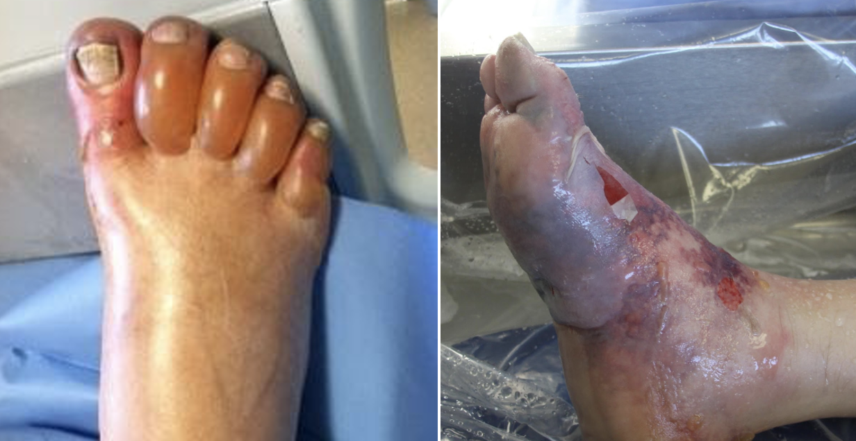
  • First-degree frostbite: Superficial damage to the skin from tissue freezing with redness (erythema), some edema, hypersensitivity, and stinging pain.

  • Second-degree frostbite: Deeper damage to the skin with a hyperemic or pale appearance, significant edema with clear or serosanguinous fluid-filled blisters, and severe pain. Frostnip, first and second-degree frostbite will generally heal without significant tissue loss.

  • Third-degree frostbite: Deep damage to the skin and subcutaneous tissue. Cold, pale, and insensate without a lot of tissue edema. Shortly after rewarming, edema rapidly forms along with the presentation of hemorrhagic blisters. Significant pain often occurs after rewarming.

  • Fourth-degree frostbite: All the elements of a third-degree injury with evidence of damage extending to the muscle, tendon, and bone of the affected area.

Figure 2. 1st and 2nd degree frostbite (left), 3rd and 4th degree frostbite (right)

Figure 2. 1st and 2nd degree frostbite (left), 3rd and 4th degree frostbite (right)

Pre-hospital or Emergency Department Management

  • Determining the extent of frostbite injury starts with a detailed history regarding how the affected area appeared on presentation.

  • The history of a cold, white, and insensate extremity on presentation is consistent with severe frostbite injury (3rd and/or 4th-degree frostbite).

  • A severe frostbite injury requires emergent therapy with thrombolytics unless the patient meets one of the exclusion criteria.

  • If in question regarding the depth of the injury, a clinical exam can be supported by a vascular study as indicated. A digital Doppler exam is a simple and quick modality to further Clarify the diagnosis of severe frostbite.

  • Complete a primary survey to rule out any traumatic injuries.

  • Correct hypothermia (warm room, remove wet clothing & jewelry, warmed fluids, etc.)

  • If there are areas of frozen tissue rapid rewarming is preferred (see next section, rapid rewarming is associated with the best outcomes and salvage rates. However, never thaw until the risk of re-freezing has been eliminated. Patients undergoing freeze-thaw cycles do not respond to thrombolytics and are treated with standard supportive frostbite therapy.

  • Protect affected areas from further trauma with padding, splinting, and immobilization while transporting.

  • Keep the patient non-weight bearing to avoid incurring additional injury to frozen tissue (ice crystals) and/or disrupting blisters.

  • Elevate the affected extremities when able to decrease tissue edema.

  • Obtain a large-bore peripheral IV & start warmed fluids. Most patients will present with dehydration secondary to hypothermia and/or intoxication.

  • Avoid direct radiant heat to prevent iatrogenic burns to the cold tissue.

  • Update the patient’s tetanus status

  • Expect the patient to have increasing pain as the involved tissue is rapidly rewarmed. Pain management should include scheduled Ibuprofen (800 mg if no contraindication) to block the arachidonic cascade, gabapentin (nerve pain), and narcotics as needed.

Figure 4. Rewarming

Figure 3. Rewarming

Rapid rewarming

  • Circulating water bath when able. Put each affected area in its own water bath to avoid the tissue “knocking” against each other.

Document start & completion time

  • Try to keep the water temp at 104 ºF (40º C)

  • It will take 30-45 min for a hand or foot

  • If the patient has boots, socks, gloves, etc frozen to the skin do not force off. Submerge the entire area as part of the rapid rewarming process

  • Continue until frostbitten limb becomes flushed red or purple, and tissue soft and pliable to gentle touch

Air Dry

  • Avoid any aggressive manipulation to decrease tissue loss and injury

  • Elevate the affected areas to decrease swelling

  • Dress the affected areas with bulky padded dressings for transfer to avoid trauma to the areas

  • Avoid rewarming with a direct heat source (heat lamp, warm IV bag, etc.). This will lead to a thermal injury secondary to the lack of blood flow.

Rewarming will be associated with:

  • A return of sensation, movement, and possible initial flushing of the skin. The vessels in the case of severe frostbite (3rd or 4th degree) quickly become thrombotic (<20 minutes) with mottling or demarcation, however, the demarcation may be subtle at first and requires careful observation.

  • In the case that the tissues return fully to a normal color and palpable pulses or Doppler digital signals are present, the patient may not need any further intervention other than close observation (inpatient or daily visits in the clinic) and pain management.

  • If any question exists, an urgent triple-phase bone scan can support perfusion to the affected area.

Figure 4. Early evidence of demarcation and patchy thrombosis

Figure 4. Early evidence of demarcation and patchy thrombosis

Indications for Thrombolytics

  • Patient presenting with frozen tissue (severe frostbite, 3rd and/or 4th degree)

  • Absent or weak Doppler pulses following rewarming

  • Clinical exam consistent with severe frostbite

  • < 24 hours of warm ischemia time (time from rewarming)

  • Time matters significantly. For each hour after rewarming delaying the start of thrombolytics decreased salvage rates even by 28.1%.

  • With correct training after discussion with a burn center that does a lot of frostbite care, thrombolytics can be safely started at the outside hospital prior to transfer to the center.

Frostbite Thrombolytic Protocol

  • Examine for any associated injuries or illnesses. If any question of injury the patient will require a head, chest, and abdominal CT to rule out any sources of bleeding.

  • The dosing of the thrombolytic requires an actual weight and while infusing the thrombolytic requires ICU status and monitoring for 24 hours.

  • Following completion of the therapy, the patient will immediately be started on treatment dose Enoxaparin for 1-2 weeks.

Figure 5. Patient before and after receiving thrombolytics

Figure 5. Patient before and after receiving thrombolytics

Contraindications to the Thrombolytic Protocol

Absolute contraindications:

  • > 24 hours of warm ischemia time

  • Repeated freeze/thaw cycles

  • Concurrent or recent (within 1 month) intracranial hemorrhage, subarachnoid hemorrhage or trauma with active bleeding

  • Inability to consistently follow a neurologic exam (eg. intubated and sedated, significant dementia)

  • Severe uncontrollable hypertension

Relative contraindications:

  • History of GI bleed or stroke within 6 mo.

  • Recent intracranial or intraspinal surgery or serious head trauma within 3 months

  • Pregnancy

Figure 6. Clinical guide for the management of frostbite

Figure 6. Clinical guide for the management of frostbite

Frostbite Take-Home Points

  1. Rapid rewarming of frozen tissue in a circulating water bath is the preferred method of rewarming.

  2. Patients that have undergone trauma in conjunction with the frostbite injury are not an absolute contraindication to receiving tPA.

  3. Starting tPA at the outside hospital, prior to transport, results in significantly improved outcomes even compared to those that receive it at UCH.

  4. Frostbite patients, regardless of whether or not they get thrombolytics, do better at a center that has experience and protocols to take care of frostbite.

Anne Wagner.png
 

Anne Lambert Wagner, MD, FACS

Associate Professor

University of Colorado

Medical Director

Burn & Frostbite Center at UC Health


How To Cite This Post…

[Peer-Reviewed, Web Publication] King, P. Porras, N. (2021, Aug 16). TPA in Frostbite. [NUEM Blog. Expert Commentary by Lamber Wagner, A]. Retrieved from http://www.nuemblog.com/blog/TPA-in-frostbite.


Other Posts You Might Enjoy


References

Cauchy E, Davis CB, Pasquier M, Meyer EF, Hackett PH. A New Proposal for Management of Severe Frostbite in the Austere Environment. Wilderness & Environmental Medicine. 2016;27(1):92-99. doi:10.1016/j.wem.2015.11.014.

Cline D, Ma OJ, Meckler GD, et al. Cold Injuries. In: Tintinalli's Emergency Medicine: a Comprehensive Study Guide. New York: McGraw-Hill Education; 2020:1333-1337.

Grayzel J, Wiley J. What’s New in Emergency Medicine. In: UpToDate, Post TW (Ed), UpToDate, Waltham, MA. (Accessed on November 3, 2020.)

Lee J, Higgins MCSS. What Interventional Radiologists Need to Know About Managing Severe Frostbite: A Meta-Analysis of Thrombolytic Therapy. American Journal of Roentgenology. 2020;214(4):930-937. doi:10.2214/ajr.19.21592.

Pandey P, Vadlamudi R, Pradhan R, Pandey KR, Kumar A, Hackett P. Case Report: Severe Frostbite in Extreme Altitude Climbers—The Kathmandu Iloprost Experience. Wilderness & Environmental Medicine. 2018;29(3):366-374. doi:10.1016/j.wem.2018.03.003.

Patel N, Srinivasa DR, Srinivasa RN, et al. Intra-arterial Thrombolysis for Extremity Frostbite Decreases Digital Amputation Rates and Hospital Length of Stay. Cardiovascular and Interventional Radiology. 2017;40(12):1824-1831. doi:10.1007/s00270-017-1729-7.

Twomey JA, Peltier GL, Zera RT. An Open-Label Study to Evaluate the Safety and Efficacy of Tissue Plasminogen Activator in Treatment of Severe Frostbite. The Journal of Trauma: Injury, Infection, and Critical Care. 2005;59(6):1350-1355. doi:10.1097/01.ta.0000195517.50778.2e.

Wagner A, Orman R. Frostbite, Asystole, Perfectionism, EQ, Middle Way, Flu. January 2019 - Frostbite - Frostbite, Asystole, Perfectionism, EQ, Middle Way, Flu | ERcast. https://www.hippoed.com/em/ercast/episode/frostbite/frostbite. Published 2019. Accessed November 3, 2020.

Zafren K, Crawford Mechem C. Frostbite: Emergency Care and Prevention. In: UpToDate, Post TW (Ed), UpToDate, Waltham, MA. (Accessed on November 3, 2020.)

Posted on August 16, 2021 and filed under Environmental.

ED Clinical Decision Making Units

Written by: Mitchell Blenden, MD (NUEM ‘24) Edited by: Em Wessling (NUEM ‘22) Expert Commentary by: Tim Loftus, MD, MBA

Written by: Mitchell Blenden, MD (NUEM ‘24) Edited by: Em Wessling (NUEM ‘22) Expert Commentary by: Tim Loftus, MD, MBA


CDU1.png
CDU2.png
CDU3.png

Expert Commentary

Thank you to Dr’s Blenden and Wessling for the excellent overview of ED CDUs including some background and indications for their use. 

 Several points to highlight and elaborate upon include the following:

The background of the utility of CDUs mostly stems from their early function as rapid diagnostic and treatment centers (RDTCs) for chest pain.  The function and utility of CDUs have since grown to demonstrate clinical benefits well-established across a variety of conditions as Dr’s Blenden and Wessling have mentioned, including not only chest pain (rule out acute coronary syndrome) but also TIA, CHF, asthma, COPD, cellulitis, pyelonephritis, pneumonia, etc. 

Value and Benefits

The utility and value of ED CDUs will continue to expand.  The percentage of all hospital admissions that start in the ED continues to grow -- 67% in 2019, up from 58% in 2004 according to the ED Benchmarking Alliance.  Additionally, EDs cared for approximately 158M people as of 2018 (EMNet/NEDI-USA), up 32% over a 10 year period.  As the number of admissions continues to grow, and considering that some of these inpatient stays are short, it follows that many of these short inpatient admissions are subject to recovery audit contractors and payor denials.  Many clinical conditions which are often subject to short inpatient stays can be cared for in dedicated short stay observation units without adversely affecting, and for the most part improving, the quality of care delivery, safety, satisfaction, cost savings, and reducing subsequent inpatient LOS.  

All stakeholders in the health care system benefit from CDU use: patients are more accurately diagnosed before leaving the ED and are discharged home faster, payors avoid costly inpatient admission charges, hospitals keep scarce inpatient bed capacity open for more appropriate patients and avoid audits and denials, and providers deliver care in a setting that more appropriately matches patient needs to resources.

Dedicated Units with Protocolized Care

Observation patients can be managed in a variety of settings and contexts, but best practice that leads to best outcomes would be in dedicated observation units adherent to protocols tailored to the patients’ conditions, the best available evidence, and local institutional resources.

Shorter hospitalizations are more likely to occur in dedicated observation units under protocols than with unstructured hospitalization on inpatient teams and simply billing status changes to observation.

Financial Considerations

Much of the existing evidence has demonstrated that CDUs can provide care that efficiently utilizes resources and results in shorter hospital lengths of stay relative to other projects to expand capacity.   Further, hospitals may realize decreased operating expenses for those patients subsequently discharged home from the CDU who have diagnoses or clinical conditions that are not as profitable for the hospital to manage in the inpatient setting - for example, CHF, which can often create a loss for the hospital.  That being said, hospitals should be careful about shifting too much acute care into CDUs, because any CDU stay that subsequently results in inpatient admission (about 20% or so) are only paid by a single DRG, which includes that care provided for in the ED, CDU, and hospital unit. Thus, you can risk incurring additional costs without additional revenue. Finally, the duration of observation should exceed 8 hours only to justify the added expense of operating the CDU, because payors, including Medicare, generally do not pay clinical or facility fees for observation stays less than 8 hours.

Another consideration when estimating value created by a CDU is the increasing use by those who would have otherwise been discharged from the ED.  It is important to consider the value of a CDU not only by the cost savings to the hospital and patient but also the possible supply-induced demand of health care services and overutilization of those services to a detriment.

CMS, and other payors, do not necessarily exclude payment from observation status patients whose stay lasted longer than 24 or even 48 hours. However, the profit margin and efficiency are reduced when patients are staying in the CDU that long, highlighting an opportunity to evaluate your particular unit’s effectiveness, efficiency, and patient selection.

Final Considerations

CDUs are not appropriate for all EDs, as only about 5-10% of ED patients have been found to be appropriate for a CDU, and in order to optimize operational and financial efficiency, a certain minimum number of beds and fixed costs would need to be overcome. 

It is worth mentioning that protocol-driven CDUs in proximity to an ED with dedicated diagnostic and treatment algorithms, patient selection criteria, predetermined outcomes and end points have demonstrated the best outcomes with respect to cost savings, patient satisfaction, safety, and reduction in hospital LOS.  For administrative and clinical operations leaders, tracking process and outcome metrics such as LOS, occupancy rate, discharge rate, and bed turns in addition to other clinical and quality outcomes will enable ongoing continuous optimization of the CDU.

Depending on the resources and throughout considerations of each hospital and health system, at times CDUs provide great benefit in being able to flexibly accommodate inpatient holds, pre or postoperative patients, or additional acute ED treatment space as the need allows.  Design and construction with this in mind may enable the hospital to best accommodate ever changing dynamics - COVID being one example.

References

  1. Emergency Medicine Network (EMNet). National Emergency Department Inventory – USA. https://www.emnet-usa.org/research/studies/nedi/nedi2018/. Accessed 1 Jan 2021.

  2. Emergency Department Benchmarking Alliance (EDBA). Before there was COVID - 2019 Emergency Department Performance Measures Report. Accessed 1 Jan 2021.

  3. Baugh CW, Liang L-J, Probst MA, Sun BC. National Cost Savings From Observation Unit Management of Syncope. Academic Emergency Medicine. 2015;22(8):934-941. doi: 10.1111/acem.12720.

  4. Baugh, C. W., Venkatesh, A. K., & Bohan, J. S. (2011). Emergency department observation units: a clinical and financial benefit for hospitals. Health care management review, 36(1), 28-37.

  5. Baugh and Granovsky - ACEP Now - https://www.acepnow.com/article/new-cms-rules-introduce-bundled-payments-for-observation-care/?singlepage=1

  6. Making Greater Use Of Dedicated Hospital Observation Units For Many Short-Stay Patients Could Save $3.1 Billion A Year. Health Affairs. 2012;31(10):2314-2323. doi: 10.1377/hlthaff.2011.0926.

  7. Ross MA, Hockenberry JM, Mutter R, Barrett M, Wheatley M, Pitts SR. Protocol-driven emergency department observation units offer savings, shorter stays, and reduced admissions. Health Aff (Millwood). 2013;32(12):2149-2156. doi: 10.1377/hlthaff.2013.0662.

  8. Rydman RJ, Zalenski RJ, Roberts RR, et al. Patient satisfaction with an emergency department chest pain observation unit. Ann Emerg Med. 1997;29(1):109-115. doi: 10.1016/s0196-0644(97)70316-0.

Tim Loftus.jpg

Timothy Loftus, MD, MBA

Assistant Professor

Department of Emergency Medicine

Northwestern University


How To Cite This Post:

[Peer-Reviewed, Web Publication] Blenden, M. Wessling, E. (2021, Apr 12). ED Clinical Decision Making Units. [NUEM Blog. Expert Commentary by Loftus, T]. Retrieved from http://www.nuemblog.com/blog/ed-clinical-decision-making-units.


Other Posts You May Enjoy

Posted on April 12, 2021 and filed under Administration.

Kawasaki Disease

Written by: Richmond Castillo, MD (NUEM ‘23) Edited by: Andra Farcas (NUEM ‘21) Expert Commentary by: Wee-Jhong Chua, MD

Written by: Richmond Castillo, MD (NUEM ‘23) Edited by: Andra Farcas (NUEM ‘21) Expert Commentary by: Wee-Jhong Chua, MD


kawasaki disease.png

Expert Commentary

A great summary of a rare, but important illness to consider in children with prolonged fevers. Kawasaki disease is a vasculitis to small and medium vessels, often occurring in childhood. While most children recover, there are important cardiac complications that need to be considered. Treatment with IVIG within the first 10 days of illness has been shown to reduce the prevalence of coronary artery aneurysms. In fact, untreated disease has been associated with an incidence of coronary artery aneurysm as high as 25%. [1]

The diagnosis of this disease process is challenging as there are no definitive testing methodologies and even the etiology of the systemic inflammatory process remains unknown. While most guidelines include 5 days of fever, the 2017 American Heart Association Scientific Statement indicates that 4 days of fever with the corresponding clinical features can be diagnostic. Moreover, in rare instances experienced clinicians have been able to make the diagnosis as early as 3 days. [1] In clinical practice, the clinical pathways at leading children’s hospitals use 4 days as a threshold for evaluation with the presence of 4 or 5 principal clinical features are present. [2,3] However, the standard practice continues to be 5 days for the classic diagnosis.

With high clinical suspicion for Kawasaki Disease, transfer to a pediatric hospital should be considered with consultations with Infectious Disease, Cardiology, and Rheumatology. It is important to have a high clinical suspicion for Kawasaki disease in children with prolonged fevers.

References

  1. McCrindle BW, Rowley AH, Newburger JW, et al. Diagnosis, treatment, and long-term management of Kawasaki disease: A scientific statement for health professionals from the American Heart Association. Circulation. 2017;135(17):e927-e999. doi:10.1161/CIR.0000000000000484

  2. D. Whitney, MD; K. Dorland, BSN; J. Beus, MD; J. Brothers, MD; L. Buckley, MD; S. Burnham MD; D. Campeggio, MSN; K. DiPasquale, MD; H. Ghanem MD; J. Hart MD; J. Lavelle, MD; C. Law PharmD; S. Natarajan, MD; J. Ronan, MD; V. Scheid, MD; S. Swami, MD; H. Ba C. Kawasaki Disease or Incomplete Kawasaki Disease Clinical Pathway — Emergency Department and Inpatient | Children’s Hospital of Philadelphia. Accessed October 20, 2020. https://www.chop.edu/clinical-pathway/kawasaki-disease-incomplete-kawasaki-disease-clinical-pathway

  3. Seattle Children’s Hospital, M Portman, M Basiaga, E Beardsley, R Engberg, K Hayward, K Kazmier, M Leu, R Migita, J Rasiah, R Sadeghian, S Sundermann, S Vora, S Zaman, 2018 February. Kawasaki Disease Pathway. Available from: http://www.seattlechildrens.org/pdf/Kawasaki-Disease-Pathway.pdf

Wee Chua.PNG

Wee-Jhong Chua, MD

Attending Physician, Pediatric Emergency Medicine

Ann & Robert H. Lurie Children's Hospital of Chicago


How To Cite This Post:

[Peer-Reviewed, Web Publication] Castillo, R. Farcas, A. (2021, Feb 1). Kawasaki Disease. [NUEM Blog. Expert Commentary by Chua, W]. Retrieved from http://www.nuemblog.com/blog/kawasaki-disease.


Other Posts You May Enjoy

Posted on February 1, 2021 and filed under Pediatrics.

Lipid Emulsion Therapy for Local Anesthetic Systemic Toxicity

Written by:&nbsp;Dana Loke, MD (NUEM ‘20)&nbsp;Edited by:&nbsp;Jim Kenny, MD (NUEM ‘18)&nbsp;Expert Commentary by: Patrick Lank, MD, MS

Written by: Dana Loke, MD (NUEM ‘20) Edited by: Jim Kenny, MD (NUEM ‘18) Expert Commentary by: Patrick Lank, MD, MS


Local anesthetic systemic toxicity (LAST) is a feared complication of local anesthetic use. Current estimates of LAST toxicity in adults range from 7.5 to 20 per 10,000 peripheral nerve blocks and 4 per 10,000 epidurals.[1] Although rare, this complication can be fatal. Unfortunately, many physicians are unaware of the toxic dose of local anesthetics and are unable to recognize the signs and symptoms of this toxicity.[2] For this reason and the fact that local anesthetic toxicity is rare, by the time this syndrome is identified, patients are often in cardiac arrest or peri-arrest. Thankfully, lipid emulsion such as Intralipid is a safe and effective therapy used to treat LAST.

 How does lipid emulsion therapy work?

Lipid emulsion therapy is an intravenous therapy that binds lipophilic toxins and therefore reverses their toxicity. There are several brand name lipid emulsion therapies, however Intralipid, a soy-based lipid emulsion that contains long-chain triglycerides, is the most commonly used (Figure 1).[3] The ability of lipid emulsion therapy to counteract the toxic effects of local anesthetics was discovered in 1998 by Weinberg et al when it was incidentally found that lab rats pre-treated with an infusion of lipids could withstand larger doses of bupivacaine before arresting.[4] The rats were also more easily resuscitated if given lipid emulsion therapy.[1]  These findings were subsequently confirmed in other laboratories and clinical systemic analyses.[5] Once studied more directly, it was found that intralipid acts as a “sink” by creating a lipid compartment within the plasma that attracts lipophilic compounds, such as local anesthetics, into the lipid sink, which is separate from the aqueous phase of the plasma.[1]

Figure 1: Composition of Various Brands of Lipid Emulsions[1]

Figure 1: Composition of Various Brands of Lipid Emulsions[1]

How does LAST manifest?

Toxicity is a rare but potentially lethal side effect of local anesthetic. However, since patients often present without any knowledge that they were administered toxic doses of local anesthetic, it is important that the EM physician be cognizant of the signs of this toxicity. Symptoms typically start after a toxic dose of local anesthetic is administered or if local anesthetic is inadvertently administered directly into a vessel instead of subcutaneously (Figure 2). Onset of LAST is typically 30 seconds to 60 minutes after administration of the anesthetic but more often than not occurs within 1-5 minutes.[6]

Figure 2: Maximum Doses and Durations of Various Local Anesthetics[9]

Figure 2: Maximum Doses and Durations of Various Local Anesthetics[9]

Symptoms of LAST can vary, however there are 5 general ways in which LAST presents.[6] One or all of these manifestations may be present.

  • CNS (excitement) – an early manifestation of LAST that often begins with confusion or slurred speech but may include subjective symptoms like metallic taste in the mouth, tinnitus, oral numbness, dizziness, lightheadedness, or visual or auditory disturbances. If not treated promptly, these symptoms often progress to seizures, syncope, coma, respiratory depression, or cardiovascular collapse.

  • Cardiovascular – often preceded by CNS symptoms but not always. May include hypertension, tachycardia or bradycardia, arrhythmias, and asystole. Depressed contractility of the heart then leads to progressive hypotension and ultimately cardiac arrest.

  • Hematologic – methemoglobinemia, cyanosis

  • Allergic – urticaria, rash, and rarely anaphylaxis

  • Local tissue response – numbness, paresthesia

The EM physician should maintain a high level of suspicion should a patient present after a same day surgery or procedure with any constellation of these symptoms.

How is lipid emulsion therapy administered?

Once LAST is recognized, the EM physician should immediately consider giving lipid emulsion therapy. An initial dose of 20% lipid emulsion at 1.5 ml/kg or a 100 ml bolus can be administered over a few minutes. This can be repeated after 5 minutes for 2 or more times for persistent hemodynamic instability. The bolus(es) should immediately be followed by a continuous infusion at 0.25-0.5 ml/kg/min.[3] The infusion should run for a minimum of 10 minutes after return of hemodynamic stability, however there are documented reports of recurrent systemic toxicity even after this. For this reason, patients should be admitted for at least 12 hours for observation and additional doses of intralipid as needed for rebound symptoms or hemodynamic compromise.[3] Consultation with your facility’s poison center is also crucial to further guide management.

Efficacy

In terms of efficacy, case reports and systemic analyses have found that lipid emulsion therapy:

  • Can reverse both neurologic and cardiac toxicity [5]

  • Leads to significantly higher rates of ROSC compared to saline controls in animal models [5]

  • Is more effective for witnessed events (for example, brief down time for patients that arrest)5

  • Is often effective in patients in which epinephrine, vasopressin, and antiarrhythmic medications did not work

Both hypoxia and acidosis worsen the toxicity of local anesthetics and may inhibit lipid emulsion therapy, so it is imperative that oxygenation and acid-base status are optimized when lipid emulsion therapy is needed.[3, 5]

 Contraindications, Complications, and Special Populations

There are no absolute contraindications to intravenous lipid emulsion therapy and no clinically significant complications documented in the literature. The benefits of lipid emulsion therapy will often outweigh any potential risks in patients with LAST, especially if hemodynamically unstable or coding.

Potential complications of lipid emulsion therapy are mainly related to hypersensitivity. Patients allergic to soybean protein or eggs theoretically may develop allergic or anaphylactic reactions. These reactions should be treated like all other allergic or anaphylactic reactions: with anti-histamines, steroids, and epinephrine as needed. Additionally, there are reported cases of hyperamylasemia however no documented progression to clinical pancreatitis.[3] There are also case reports of extreme lipemia, however even a patient that was inadvertently given 2 L of 20% lipid emulsion did not develop any cardiopulmonary complications.[5] The lipemia however did interfere with standard laboratory tests.[5]

Intralipid is safe in pregnancy and has documented use for treating LAST in term pregnancy.[7] Furthermore, it has documented uncomplicated use in pediatric and neonatal patients.[3, 8]

 Key Points

  • Systemic toxicity is a rare but potentially fatal complication of local anesthetic use.

  • Lipid emulsion therapy such as Intralipid mitigates the toxic effects of local anesthetics and can reverse both neurologic and cardiac toxicity.

  • LAST may manifest initially with CNS symptoms but can progress to seizure, respiratory depression, coma, and cardiovascular collapse.

  • An initial bolus of 1.5 ml/kg or 100 ml 20% lipid emulsion followed by an infusion starting at 0.25 ml/kg/min is crucial to reverse toxicity and prevent recurrence.

  • Hypoxia and acidosis both worsen LAST and may inhibit lipid emulsion therapy.

  • Patients should be admitted in order to monitor for recurrent toxicity.

  • There are no contraindications to and minimal side effects of lipid emulsion therapy.


Expert Commentary

Thank you both for the above thorough review of local anesthetic systemic toxicity (LAST) from the emergency physician perspective! I only want to add a few points to consider when learning more about LAST.

Without going into too much detail, there has been a lot of research done to figure out exactly how lipids aide in the treatment of patients with severe LAST. The lipid sink model is wonderfully understandable and explains many of the clinical and laboratory we see (e.g., a greater decrease in free serum concentration of more lipophilic local anesthetics).  However, there are some other models and theories to be aware of. One I am fascinated by is the “lipid shuttle.” Fundamentally, this describes the phenomenon that lipid therapy will decrease the concentration of local anesthetic at sites of toxicity (i.e., heart and CNS) and increase its concentration in the liver. So instead of lipids acting only as a “sink” to remove a toxin from free availability, it is helping mobilize the toxin to an area where it can go through the process of elimination from the body. Additionally, there are wonderful biochemical explanations (e.g., fatty acid supply, inhibition of nitric oxide release, reversal of mitochondrial dysfunction) to the positive cardiovascular effects seen after lipid treatment in LAST. All of these explanations, it seems, combine to contribute to the hemodynamic response seen in LAST.

Second, I would like to point your readers towards a resource that may help them work through the mechanics of administering lipid rescue therapy in LAST – lipidrescue.org. On that website, one can find links to various protocols, compilations of prior research done on the topic, and much more background on the science of the treatment than I provided above.

Third, in the emergency department, I think you are correct in saying that the most likely source of LAST we would see would come from outpatient surgery centers. A few other clinical scenarios to be aware of would include the following: ingestion of local anesthetics – mostly benzonatate (Tessalon); non-surgical outpatient aesthetic offices that may use topical anesthetics; inappropriate and excessive home use of local anesthetics for pain relief.

Finally, a very brief comment on the use of lipid rescue therapy in non-LAST toxic exposures although that was not the subject of your post. While lipid rescue therapy for LAST has a remarkable record of being effective, that is not yet the case with its use in other toxic exposures. A list of the side effects of lipid rescue therapy includes but is not limited to ARDS, pancreatitis, infection, and significant laboratory interference. While in the setting of severe LAST, the risk: benefit often favors administering lipid rescue, this may not be the case in the setting of non-LAST exposures.  For those non-LAST cases (as well as with LAST cases) in which you are wondering if lipid rescue would be appropriate, I would strongly recommend you call your regional poison center to discuss further focused therapy. 

Patrick_Lank-04 (1).jpg

Patrick Lank, MD, MS

Assistant Professor of Emergency Medicine

Medical Toxicologist

Department of Emergency Medicine


How To Cite This Post:

[Peer-Reviewed, Web Publication] Loke D, Kenny J. (2020, July 20). Lipid Emulsion Therapy for Local Anesthetic Systemic Toxicity. Expert Commentary by Lank P. Retrieved from http://www.nuemblog.com/blog/lipid-emulsion-therapy


Other Posts You May Enjoy


References

1.     Manavi, M. (201). Lipid infusion as a treatment for local anesthetic toxicity: a literature review. AANA Journal, 78(1), 69-78.

2.     Cooper, B.R., Moll, T., & Griffiths, J.R. (2010) Local anaesthetic toxicity: are we prepared for the consequences in the Emergency Department. J Emerg Med, 27(8), 599.

3.     Mercado, P. & Weinberg, G.L. (2011). Local anesthetic systemic toxicity: prevention and treatment. Anesthesiology Clin, 29(2), 233-242.

4.     Weinberg, G.L., VadeBancouer, T., Ramarju, G.A., Garcia-Amaro, M.F., & Cwik, M.J. (1998). Pretreatment or resuscitation with a lipid infusion shifts the dose-response to bupivacaine-induced asystole in rats. Anesthesiology, 88(4), 1071-5.

5.     Weinberg, G.L. (2012). Lipid emulsion infusion: resuscitation for local anesthetic and other drug overdose. Anesthesiology, 117(1), 180-7.

6.     Wadlund, D. (2017). Local anesthetic systemic toxicity. ARON Journal, 106(5), 367-77.

7.     Dun-Chi Lin, J., Sivanesan, E., Horlocker, T.T., & Missair, A. (2017). Two for one: a case report of intravenous lipid emulsion to treat local anesthetic systemic toxicity in term pregnancy. A&A Case Reports, 8(9), 235-7.

8.     Shah, S., Gopalakrishnan, S., Apuya, J., Shah, S., & Martin, T. (2009). Use of intralipid in an infant with impending cardiovascular collapse due to local anesthetic toxicity. J Anesth, 23(3), 439-441.

9. “Missouri Society of Health-System Pharmacists - Overview of Management of Local Anesthetic Systemic Toxicity (LAST) Based on Updated 2017/18 ASRA Practice Guidelines.”

Posted on July 20, 2020 and filed under Toxicology.

Mood Stabilizer Toxicities

Written by: Justine Ko, MD (NUEM PGY-4) Edited by: Sarah Dhake, MD (NUEM ‘19) Expert Commentary by: Patrick Lank, MD, MS

Written by: Justine Ko, MD (NUEM PGY-4) Edited by: Sarah Dhake, MD (NUEM ‘19) Expert Commentary by: Patrick Lank, MD, MS


mood-stabilizer_36306943.png

Expert Commentary


This is a great summary of the causes, symptoms, work-up, and treatment of two relatively common medications that cause toxicity. In fact, these (along with carbamazepine) are levels I routinely recommend checking in patients with a history of bipolar disorder who come to the emergency department with altered mental status even if they do not report a history of being on these medications. That is because these three medications are very commonly used in the treatment of bipolar disorder and all have quite different treatment courses. In addition to the great summary above, below are some of my usual teaching points about these medications in overdose.

Let's tackle them separately as they are quite different toxicities.

First let's talk about lithium. In almost all medical texts, the tissue distribution of lithium is appropriately identified as being "complex." The easiest way I communicate that with patients, families, and medical learners is that in chronic therapy, lithium forms "stores" of drug in the body and intracellularly. Clinically that is relevant because after performing hemodialysis (HD) for lithium toxicity, you will reliably see an initial drop in lithium concentration followed by elevation approaching pre-dialysis levels if routine HD is performed. Although that could make one feel ambivalent about routinely recommending HD for lithium toxicity, there is suggestion of an alternate advantage of performing HD for lithium toxicity.  Vodovar et al published a study in 2016 showing that patients who met their institutional criteria for HD and had HD performed had significantly fewer neurologic side effects from their toxicity than those who met criteria but did not have HD. So even though it did not impact usual measurements that we would expect HD to influence -- mortality and ICU length of stay -- its performance in this study seems to have been clinically beneficial.

 The other big thing to discuss with lithium toxicity is that there are many known medication interactions with lithium. In short, any medications that impair renal function should not be used in someone on chronic lithium therapy. The main list of those medications includes NSAIDs, ACE inhibitors, ARBs, and thiazide diuretics.

 Most of the unique aspects of valproate toxicity focus on its diagnosis and treatment. In the setting of acute intentional overdose of valproate, one of the most important things for emergency physicians to be aware of is that there can be a delay of peak valproic acid level. There are cases of patients presenting to an emergency department with stated valproate ingestion, initial negative level, then repeat level hours later being in the toxic range. So I recommend serial valproate levels until both down-trending and non-toxic. For treatment, there is a great summary of recommendations by the Extracorporeal Treatments in Poisoning Workgroup (EXTRIP) published online (https://www.extrip-workgroup.org/valproic-acid). In short, consult nephrology for dialysis if the patient is super sick.

 As always, I recommend you consult your regional poison center when you are worried your patient is experiencing medication toxicity. But I hope this infographic and some of my comments helps you understand their recommendations.

 References

  1. Vodovar V, et al. Lithium poisoning in the intensive care unit: predictive factors of severity and indications for extracorporeal toxin removal to improve outcome. Clin Tox (Phila) 2016 Sep; 54(8): 615-23.

  2.  Lank P and Bryant S. "Valproic Acid" In: Wolfson A, Hendey G, Ling L, Rosen C, Schaider J, Cloutier R (eds.): Harwood-Nuss’ Clinical Practice of Emergency Medicine, 6th edition. Lippincott Williams &amp; Wilkins 2014.

Patrick_Lank-04.jpg
 

Patrick Lank, MD, MS

Assistant Professor of Emergency Medicine

Medical Toxicologist

Department of Emergency Medicine


 How To Cite This Post

[Peer-Reviewed, Web Publication] Ko J, Dhake S. (2020, July 13). Mood Stabilizer Toxicities [NUEM Blog. Expert Commentary by Lank P. Retrieved from http://www.nuemblog.com/blog/mood-stabilizer-tox


Other Posts You May Enjoy

Posted on July 13, 2020 and filed under Toxicology.

Marathon: The Collapsed Athlete

marathon image.png

Written by: Zach Schmitz, MD (PGY-3)  Edited by: Andrew Berg, MD (NUEM ‘19)  Expert Commentary by: Jake Stelter, MD


Marathon: The Collapsed Athlete

You’ve been enjoying a beautiful, 71 degree day wrapping ankles and rehydrating runners at the last marathon medical tent, just one mile from the finish. Suddenly, you get a different, more concerning type of call - there’s a runner down about a block south.

You fight against the flow of runners and finally see your patient on the left side of the course. He’s laying on his back and a bystander has placed an ice bag on his head. He tells you his name is Tony, but can’t tell you where he is or what he was doing. A friend says he was slowing down and looking unsteady before sitting on the curb. He’s sweating, working a little hard to breathe, and he has a 2+ radial pulse. What is your approach?

What to rule out first:

Just as with other ED patients, the first thing to do is rule out or intervene on life-threatening causes of runner’s collapse. Collapse during exercise is particularly concerning. There are five main causes of downed runners in that category: Sudden Cardiac Arrest, Exertional Heat Stroke, Anaphylaxis, Hypoglycemia, and Hyponatremia [1]. Below is an approach aimed toward addressing these concerns.

Marathon Algorithm.png

1. Sudden Cardiac Arrest

  • Suggested by an absent pulse and/or abnormal respirations.

  • Do not delay treatment. Start ACLS/BLS as your training and equipment allows and transport to a nearby ED.

  • Extremely rare[2]

2. Anaphylaxis

  • Suggested by any combination wheezes/stridor, shortness of breath, swelling, skin changes, nausea/vomiting, and altered mental status.

  • Treatment will likely be limited to IM epinephrine, as antihistamines, H2 blockers, and steroids are not routinely stocked in medical tents.

3. Exertional Heat Stroke

  • Suggested by altered mental status and a rectal temperature of > 104 degrees F.

  • These patients should be placed in an ice bucket immediately – any delay will risk permanent neurologic dysfunction.

  • Ice bucket immersion has been shown to reduce core body temperature 3x faster than ice towels and 15x faster than ice packs over major arteries[3].

  • Rapid on-site cooling is associated with better outcomes than immediate transfer to an emergency department for cooling. Those on site cooling end-points are controversial, but getting below 102 degrees F consistently leads to a safe transfer.[4]

  • You have to use rectal temperatures, as other temperature measurements have proven unreliable in a marathon setting.[4}

4. Hypoglycemia

  • Suggested by a spectrum from tremor, anxiety, diaphoresis, and altered mental status, up to seizure and coma.

  • Patients should be treated with glucose and transferred to a nearby medical facility.

5. Hyponatremia

  • Suggested by paresthesias, nausea/vomiting, and altered mental status, up to seizure and coma.

  • In one study of the Boston Marathon, 13% of runners had sodium values < 130, and 0.6% had critical values < 120. Those with longer race times, weight gain during the race, and those at the extreme ends of the BMI scale were more likely to have problems.[5]

  • Normal saline should be started for patients with initial Na of 130 or below, and 3% NS may be considered if Na < 125.


Thankfully, the above conditions comprise the minority of visits to medical tents at the marathon (including for downed runners). So what do you do with someone who is down and lightheaded but with a temperature of 99.3, sodium of 138, glucose of 98, and no signs of anaphylaxis? Collapse during exercise is still concerning, even after ruling out the causes above. You’ll want to confirm the patient can tolerate oral rehydration, place in a Trendelenberg position, and likely refer for further testing.

Collapse after exercise is more common, and, fortunately, often benign. Exercise associated collapse is likely to be the most frequent condition you encounter if you are in the final medical tent.


Exercise Associated Collapse (EAC)

Although considered to be more a chief complaint than diagnosis, EAC is defined as “a collapse in conscious athletes who are unable to stand or walk unaided as a result of light headedness, faintness and dizziness or syncope causing a collapse that occurs after completion of an exertional event.”[4] In one study, it accounted for 59% of patient presentations at the final medical tent.[1]

While running, increased oxygen demand by muscle leads to increased cardiac output and decreased peripheral vascular resistance. Skeletal muscle works as “second heart” for the race, overcoming this decrease in PVR to increase venous return. This mechanism is lost when running stops, and blood pools in the lower extremities. Cardiac output cannot be maintained, and perfusion is decreased. Further, the baroreceptor reflex controlling this mechanism is often compromised during long exercise. [6]

It is a fairly simple mechanism to reverse, and therefore a simple condition to treat. Placing the patient in a Trendelenberg position with the legs above the heart will usually achieve a fluid equilibrium in 10-30. Holtzhausen showed that these patients have no different electrolyte concentrations and are no more volume depleted than runners who finished the race without complication, so IV fluids are unnecessary[7]. However, keep in mind these people just ran a marathon, so they could probably use a little oral rehydration.

These patients should prove capable of sitting, then standing, then walking and eating before being discharged from the tent. If they show signs of altered mental status, vital sign abnormalities, or electrolyte imbalances, they should be treated appropriate and then transferred to an emergency department.


The vast majority of runners visiting a marathon medical tent are fully capable of finishing the race and just need help working out a cramp, covering up a blister, or grabbing some gel to cool a sore muscle. However, serious conditions do happen, and it is important to keep them in mind the next time you volunteer at your local marathon.


Take away points:

  • Life-threatening pathology is certainly possible in this relatively healthy cohort

  • Sudden cardiac arrest, exertional heat stroke, anaphylaxis, hypoglycemia, and hyponatremia should be considered for every down runner with altered mental status

  • Approach to the down runner: make sure you don’t need ACLS on scene > transfer to tent for rectal temp > Na, Glucose

  • If rectal temp is > 104, go directly to ice bath. Fully cool before transferring patient from tent

  • Although syncope post race can be scary, EAC is likely to resolve with 10-30 minutes of raised legs and oral rehydration


Expert Commentary

This is a great review of managing marathon runners who are acutely ill. It is important to keep in mind the diagnoses pointed out when dealing with a collapsed athlete.

1. Sudden Cardiac Arrest: This should be treated promptly following BLS/ACLS protocols. In this situation, the goal is to get to early defibrillation if possible as the most common cause is going to be a shockable arrhythmia, either ventricular fibrillation or ventricular tachycardia. The resources immediately available to you will vary depending on where on the course the patient goes down. Early activation of EMS is critical as they will bring with them both the means of transportation as well as ACLS supplies to aid in resuscitation.

2. Altered Mental Status: In a marathon athlete, the most important and life-threatening cause of altered mental status that needs to be ruled out is exertional heat stroke. As correctly pointed out, a core rectal temperature should be obtained on any athlete that is altered. Once identified as having a core temp over 103F, the athlete should be immediately cooled in an ice water tub until their temperature is 102F. At this point, the athlete should be removed from the water. Cooling below 102F can cause rebound hypothermia as cool peripheral blood shunts to the core. Avoid starting IV’s in runners prior to cooling, as getting blood into the tubs will contaminate them. If the athlete is normothermic and altered, check for hypoglycemia and treat accordingly.

3. Hyponatremia: Exercise-Induced Hyponatremia (EIN) is a relatively rare but very serious complication of endurance events. It is generally caused by excess sodium loss (sweating) that is often accompanied by excess free water intake. In a patient that is having signs and symptoms of neurologic dysfunction that is normothermic and not hypoglycemic, consider EIN. Common signs and symptoms include paresthesias, confusion, muscle weakness, cramping and seizures. If you have the ability to check a rapid sodium, then you can treat accordingly. If the patient’s sodium level is below 130 WITHOUT neurological symptoms, restrict free water intake and consider oral rehydration with electrolyte solutions. Sparingly administer isotonic IV fluids, no more than 250-500mL at a time and recheck the sodium level after each small bolus. If a patient falls into the category of hypervolemic hyponatremia, they may actually have an excess of ADH hormone and giving fluid may precipitate an even further drop in sodium. If a hyponatremic patient is having any neurological manifestation, especially seizures, the treatment is administration of 3% sodium chloride solution in 50-100mL boluses.

Also, as pointed out, be sure to consider other potential causes of your patient’s symptoms, including but not limited to cardiac pathology, trauma, stroke, and exercise associated collapse. Patients that are undifferentiated will often need to be transported to the nearest Emergency Department, as you are unlikely to have the resources to complete a diagnostic work-up in your course medical tent.

Jake Stelter.png

Jacob Stelter, MD

Instructor of Clinical Emergency Medicine

Primary Care Sports Medicine Fellow

University of Cincinnati

Medical Committee - Lead ICU Tent Coordinator

Bank of America Chicago Marathon


How to Cite This Post

[Peer-Reviewed, Web Publication] Schmitz Z, Berg, A. (2020, April 20). Marathon: The Collapsed Athlete. [NUEM Blog. Expert Commentary by Stelter, J]. Retrieved from http://www.nuemblog.com/blog/marathon


Other Posts You Might Enjoy:


References

[1] Roberts W, O’connor F, Grayzel J. Preparation and management of mass participation endurance sporting events. UpToDate. May 23 2017. https://www.uptodate.com/contents/preparation-and-management-of-mass-participation-endurance-sporting-events

[2] Roberts W.O., and Maron B.J.: Evidence for decreasing occurrence of sudden cardiac death associated with the marathon. J Am Coll Cardiol 2005; 46: pp. 1373-1374

[3] Casa D et al. Exertional Heat Stroke: New Concepts Regarding Cause and Care. Curr Sports Med Rep. 2012 May-Jun;11(3):115-23.

[4] Childress MA, O'Connor FG, Levine BD. Exertional collapse in the runner: evaluation and management in fieldside and office-based settings. Clin Sports Med. 2010 Jul;29(3):459-76. doi: 10.1016/j.csm.2010.03.007.

[5] Almond et al. Hyponatremia among runners in the Boston Marathon. N Engl J Med. 2005 Apr 14;352(15):1550-6.

[6] Asplund CA, O'Connor FG, Noakes TD Exercise-associated collapse: an evidence-based review and primer for clinicians Br J Sports Med 2011;45:1157-1162.

[7] Holtzhausen LM1, Noakes TD, Kroning B, de Klerk M, Roberts M, Emsley R. Clinical and biochemical characteristics of collapsed ultra-marathon runners. Med Sci Sports Exerc. 1994 Sep;26(9):1095-101.

[8] Madan S., Chung E., The Syncopal Athlete. American College of Cardiology. http://www.acc.org/latest-in-cardiology/articles/2016/04/29/19/06/the-syncopal-athlete. Apr 29 2016. Accessed 10 1 2017.

Posted on April 20, 2020 and filed under Environmental.

Malingering in the Emergency Department

Malingering in the ED.png

Written by: Aaron Wibberley, MD (PGY-2)  Edited by: Kaitlin Ray, MD (PGY-4)  Expert commentary by: Chris Lipp, MD


Malingering.png

Expert Commentary

Malingering is a patient behaviour with a profound hazard: the misdiagnosis of a “deceptive” patient who in reality has a serious medical illness. Chief complaints associated with malingering may coincide with a vast differential of possibilities: neck pain, symptoms after head trauma, and abdominal pain. Just like musculoskeletal back pain is a diagnosis of exclusion for a patient presenting with acute discomfort, malingering can be considered when a patient has been thoroughly assessed based on their history and physical examination (with appropriate diagnostic testing). Tools exist to help psychiatrists, neurologists and occupational physicians in diagnosing malingering, but these are largely out of the skill set of most emergency physicians. To determine if malingering should be suspected there are several questions to consider: are there any rewards the patient may be seeking after? What incentive may the patient have to seek after hospitalization, time off work, or addictive medication prescriptions? In most cases a team-based approach involving interdisciplinary professionals and sufficient collateral information are required to (1) make the diagnosis of malingering substantiated and (2) free from the excessive medicolegal risks of misdiagnosis. Emergency department clinicians must vigilantly consider malingering, factitious disorders, and other psychiatric illness as diagnoses of exclusion.

Chris Lipp.png
 

Chris Lipp, MD

Attending Physician

Calgary Emergency Medicine

Author at CanadiEM

Co-Founder of CRACKCast


How To Cite This Post

[Peer-Reviewed, Web Publication] Wibberley, A. Ray, K. (2020, Feb 24). Malingering in the ED. [NUEM Blog. Expert Commentary by Lipp, C]. Retrieved from http://www.nuemblog.com/blog/malingering


Other Posts You Might Enjoy…

Posted on February 24, 2020 and filed under Psychiatry.