Posts tagged #tca

Bicarb in Cardiac Arrest

Written by: Kishan Ughreja, MD (NUEM ‘23) Edited by: Sean Watts, MD (NUEM ‘22)
Expert Commentary by: Dana Loke, MD (NUEM ‘21)


Utility of Sodium Bicarbonate in Cardiac Arrest

Use of sodium bicarbonate as empiric therapy in cardiac arrest has been an area of controversy.  During cardiac arrest hypoxia and hypoperfusion results in severe metabolic acidosis and subsequent impaired myocardial contractility, decreased efficacy of vasopressors, and increased risk of dysrhythmias. Previous ACLS guidelines recommended use of sodium bicarbonate to mitigate these effects; however,  harms are also associated with its routine use  including compensatory respiratory acidosis, hyperosmolarity, increased vascular resistance, and reduction in ionized calcium. 1 Current guidelines no longer recommend routine use of sodium bicarbonate, except in cases of arrest secondary to hyperkalemia, TCA overdose or preexisting metabolic acidosis.2 Regardless of these recommendations, sodium bicarbonate continues to be utilized during routine management of cardiac arrest, and studies are limited in investigating its appropriate use.

The study below investigates the effect of sodium bicarbonate in patients suffering out-of-hospital cardiac arrest with severe metabolic acidosis during prolonged CPR.


Article

Clinical Question

In patients with prolonged, atraumatic out-of-hospital cardiac arrest (OHCA) and severe metabolic acidosis, does sodium bicarbonate (SB) administration with transient hyperventilation improve acidosis without increased CO2 burden, enhance rates of return of spontaneous circulation (ROSC), survival to admission, and favorable neurologic outcomes?

Study Design

Double-blind, prospective, randomized, placebo-controlled, single-center pilot clinical trial 

Population

Inclusion criteria: Atraumatic arrest in patients ≥18yo without ROSC after 10 minutes of CPR in ED and with pH <7.1 or bicarbonate <10 mEq/L on ABG

Exclusion criteria: DNR, ECPR, ROSC w/i 10 minutes of ACLS, absence of severe metabolic acidosis on ABG after 10 minutes of CPR

Data collection over 1 year at Asan Medical Center, a tertiary referral center in Seoul, Korea

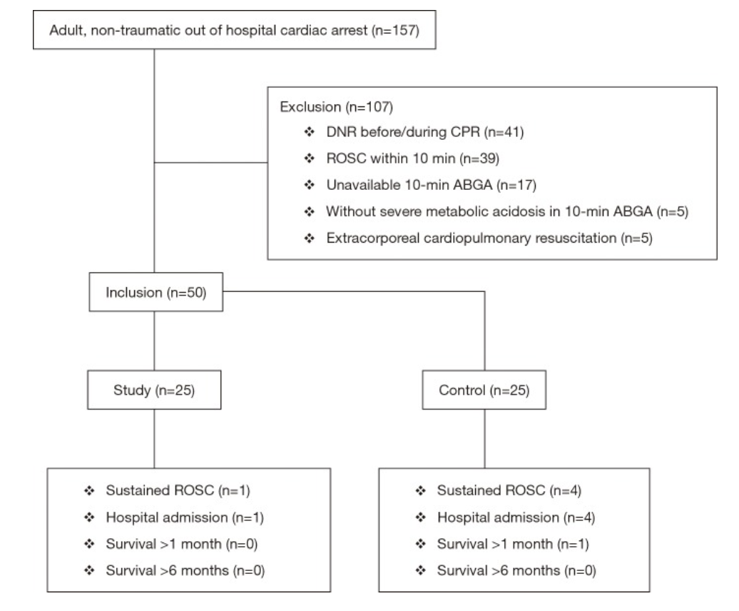
Figure 1: Patient Selection

Intervention

Sodium bicarbonate administration of 50 mEq/L over 2 minutes with concurrent increase in ventilation rate from 10 to 20 breaths per minute for 2 minutes

Control

Normal saline administration of 50 mL over 2 minutes (with same transient hyperventilation)

Outcomes

Primary

  • Change in acidosis (per methods section)

Secondary

  • Sustained ROSC — defined as restoration of a palpable pulse ≥20 min (per methods section, but listed as primary outcome in abstract)

  • Survival to hospital admission

  • Good neurological survival at 1 and 6 months (defined as cerebral performance category 1 or 2)

Results

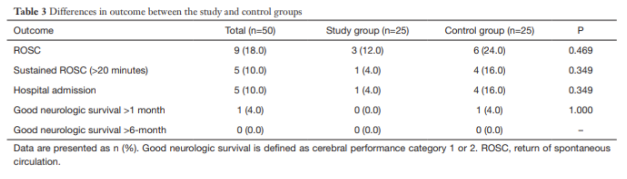
  • 157 patients presented with cardiac arrest, 50 enrolled per inclusion criteria

  • No significant differences between study and control groups regarding demographics, PMH, witnessed arrest, bystander CPR, pre-hospital and initial cardiac rhythm

  • 10% (n=5) of enrolled patients with sustained ROSC and admitted

  • No patients survived at 6 months follow up

Pre-Intervention

  • ABG results at 10 minutes were not significantly different between groups
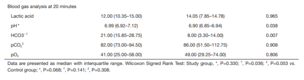
Post-intervention

  • ABG results at 20 minutes demonstrate that pH and HCO3- were higher in the study group than in the control group

    • pH 6.99 vs 6.90, p=0.038

    • HCO3- 21.0 vs 8.00, p=0.007

  • Within the study group, the increase in pH was not statistically significant after sodium bicarbonate administration; the increase in HCO3- was statistically significant (using Wilcoxon signed rank test)

  • No statistically significant findings in the control group after normal saline administration

  • No significant differences in any secondary outcomes (sustained ROSC, survival to admission, good neurologic outcome)

Strengths

  • Randomized, double-blinded, placebo-controlled study design

  • This study adds additional information to a clinical question that has limited previous research

  • This study added a practical clinical intervention (hyperventilation) to counteract excessive CO2 accumulation secondary to sodium bicarbonate administration, a known deleterious effect of this compound.

  • Strong control over sodium bicarbonate administration (no pre-hospital administration allowed in South Korea), so authors could control when it was given and analyze ABG results at desired intervals)

Weaknesses 

  • Small, single-center study with only 50 enrolled patients

  • Primary endpoint unclear from abstract vs methods, whether it was change acidosis or sustained ROSC; however, neither is truly patient-centered clinical outcome (good neurological outcome would be the ideal primary outcome)

  • Dosing was universal — 50 mEq/L instead of weight based (1-2 mEq/L/kg), which could result in improper dosing

  • Hyperventilation strategy may have benefited sodium bicarbonate administration group by countering respiratory alkalosis, however, it could have harmed the placebo group

  • Possible venous sampling rather than arterial for blood gas analysis at 10-minute point, though this would be a concern in any arrest setting if an arterial line could not be established in this time frame

Author’s Conclusion

“The use of sodium bicarbonate during CPR with transient hyperventilation improves acid-base status without CO2 elevation which is one of the most concerned adverse effects of sodium bicarbonate administration, but it had no effect on the improvement of the rate of ROSC and good neurologic survival.  At this point, we could not advise for or against its administration, our pilot data could be used to help design a larger trial to verify the efficacy of sodium bicarbonate.”

Bottom Line

Based on this study, the use of sodium bicarbonate does not appear to improve clinically significant outcomes, though it improved acid-base status.  Sodium bicarbonate should not be indiscriminately used in all cardiac arrests, and larger trials should be performed to further evaluate its impact on patient-centered outcomes.

Citation

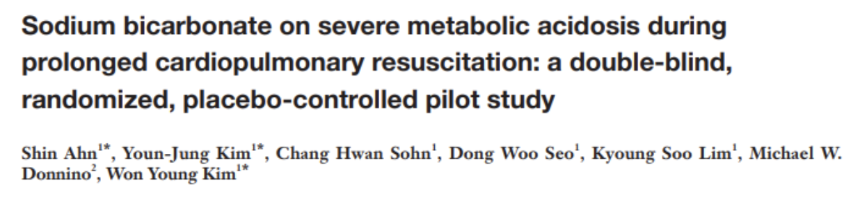
Ahn, S., Kim, Y. J., Sohn, C. H., Seo, D. W., Lim, K. S., Donnino, M. W., & Kim, W. Y. (2018). Sodium bicarbonate on severe metabolic acidosis during prolonged cardiopulmonary resuscitation: a double-blind, randomized, placebo-controlled pilot study. Journal of thoracic disease, 10(4), 2295.

References

  1. White, S. J., Himes, D., Rouhani, M., & Slovis, C. M. (2001). Selected controversies in cardiopulmonary resuscitation. Seminars in respiratory and critical care medicine, 22(1), 35–50. https://doi.org/10.1055/s-2001-13839

  2. Merchant, R. M., Topjian, A. A., Panchal, A. R., Cheng, A., Aziz, K., Berg, K. M., Lavonas, E. J., Magid, D. J., & Adult Basic and Advanced Life Support, Pediatric Basic and Advanced Life Support, Neonatal Life Support, Resuscitation Education Science, and Systems of Care Writing Groups (2020). Part 1: Executive Summary: 2020 American Heart Association Guidelines for Cardiopulmonary Resuscitation and Emergency Cardiovascular Care. Circulation, 142(16_suppl_2), S337–S357. https://doi.org/10.1161/CIR.0000000000000918


Expert Commentary

Thank you Dr. Ughreja and Dr. Watts for this excellent blog post on an important topic. In medicine, we often ask “what else can we do?” but less often do we ask “is what we’re already doing effective?” This is especially important for resuscitation and cardiac arrest. Not everything that is standard-of-care is ultimately effective care, and overtreating patients can lead to other untoward effects. 

In addition to the points made in the above blog, I would add a few important notes into the equation. First, the study excluded in-hospital cardiac arrest and therefore should not be considered in those patients. Second, the study also excluded those patients with early ROSC and absence of severe metabolic acidosis, effectively biasing towards inclusion of sicker patients. It is unclear how administration of sodium bicarbonate may have influenced those patients. Third, the study population was quite small and a striking majority of that population were found to have an initial rhythm of asystole. Fourth, ventilation rates were purposefully increased during bicarb administration. Though this may be practical and can potentially counteract excessive CO2 accumulation secondary to sodium bicarbonate administration, this is not common practice which leads to questions of this study’s external validity at other institutions.  

So, despite this study, at this point in time we still must grapple with the “should-we-or-should-we-not” of sodium bicarbonate administration in prolonged cardiac arrest. Some scenarios certainly do require sodium bicarbonate, most notably TCA overdose and hyperkalemia. In these cases, it’s obvious what to do. But so often what we do in emergency medicine is riddled with uncertainty. An unclear cause of cardiac arrest is certainly one of those situations. Perhaps instead of mindlessly giving sodium bicarbonate to cardiac arrest patients, we should give it once or twice and look for evidence that it has had an effect. Is the rhythm narrowing? Did you obtain ROSC shortly after administration? If not, giving dose after dose of sodium bicarbonate in hopes of meaningful recovery may not be the best path forward.

Dana Loke, MD

Department of Emergency Medicine

Northwestern University Feinberg School of Medicine

Northwestern Memorial Hospital


How To Cite This Post:

[Peer-Reviewed, Web Publication] Ughreja, K. Watts, S. (2021, Dec 6). Bicarb in Cardiac Arrest. [NUEM Blog. Expert Commentary by Loke, D]. Retrieved from http://www.nuemblog.com/blog/bicarb-arrest


Other Posts You May Enjoy

Posted on December 6, 2021 and filed under Critical care.

Serotonin Syndrome

Screen Shot 2017-11-11 at 12.28.34 PM.png

Written by: Jacob Stelter, MD (NUEM PGY-3) Edited by: Elizabeth Byrne, MD,  (NUEM Graduate 2017) Expert Commentary by: Patrick Lank, MD


The Case

A 21-yearold male with history of PTSD and depression presented to the ED complaining of “feeling anxious.” When evaluated, the patient was found to be anxious, continuously pacing around the room. His speech was pressured as he tangentially answered questions.  During the interview he stated that he was discharged a few days ago from a psychiatric hospital after he had intentionally overdosed on clonazepam. He then admited that two hours prior to arrival in your ED he took 30 pills of his prescribed sertraline 100mg tabs to “calm his nerves.” He denied any concurrent substance abuse. Despite the stated overdose, he denied any suicidal intent with his ingestion.


Physical Exam:

Vital Signs: T 98.9F oral   HR 137   BP  152/89   RR 20    Sat 99% on room air

General: Awake, alert, anxious-appearing, pacing around the room, mildly diaphoretic

Head: normocephalic, atraumatic

HEENT: PERRL,  EOMI, anicteric sclera, mydriatic pupils b/l, 8mm

Cardiac: tachycardic,  regular rhythm, no MRG

Resp: clear to auscultation, nonlabored, no crackles appreciated

Abd: Soft, not distended, not-tender to palpation, +bowel sounds

Extremities: well perfused, 2+ radial and DP pulses b/l, mildly diaphoretic

Neuro: AAOx3, 5/5 strength in all 4 extremities, +ankle clonus, hyperreflexia on patellar reflexes.

Psych: Cooperative but anxious with pressured and tangential speech., no HI , SI, hallucinations/delusions


Pertinent Labs:

WBC 12.5, Hgb 15.8, electrolytes within normal limits,  LA of 1.7.

Urine drug screen was positive for amphetamines and benzodiazepines. Acetaminophen, salicylate, and ethanol concentrations were below detection limits.


ECG:

Sinus tachycardia, QTc 457, no abnormal morphology of the QRS complexes


Diagnosis?

Serotonin Syndrome due to SSRI overdose.


Serotonin Syndrome

Overview

Serotonin syndrome is a disorder that is precipitated by excess serotonin. It is classically described as having a combination of hyperautonomic hemodynamic changes, neuromuscular derangements, and a change in the patient’s mental status. [1]  Serotonin syndrome is a medical condition seen only in patients with exposure to serotonergic medications and has been more recently appreciated since the advent of serotonergic drugs used to treat depression and anxiety. However, in addition to pharmaceuticals, botanicals and recreational drugs of abuse can also be serotonergic.  Causative substances increase the amount of circulating serotonin in the bloodstream, which leads to an increased propensity to develop serotonin syndrome [2].  This is not a rare diagnosis; there are approximately 7,300 cases per year results in about 100 deaths [1].

Clinical Presentation

Boyer EW, Shannon M. The serotonin syndrome. N Engl J Med 2005;352:1112-20.

The diagnosis of serotonin syndrome will often manifest around the time of a dose increase, addition of another serotonergic agent or an overdose on a serotonergic agent [3]. Serotonin syndrome will typically manifest within 24 hours of exposure to the serotonergic agent [4]. Symptom presentation  can be separated into three main categories: cognitive, autonomic and neuromuscular. Cognitive symptoms range from restlessness and anxiety to agitation and altered mental status [2].  Autonomic signs and symptoms include tachycardia, mydriasis, hyper- or hypotension, hyperthermia and diaphoresis [2]. Neuromuscular symptoms are also usually observed, ranging from akathisia to muscle rigidity, hyperreflexia and myoclonus [2].  Serotonin syndrome can be a life-threatening condition if not promptly recognized and treated as it can progress to seizures or shock [1]. 

Boyer EW, Shannon M. The serotonin syndrome. N Engl J Med 2005;352:1112-20.

Treatment

The most important initial step to the treatment of serotonin syndrome is to remove exposure to the offending agent. Use of activated charcoal to assist with gastrointestinal decontamination can be considered in the setting of a recent ingestion [1].  Following this, the cornerstone of therapy of serotonin syndrome is symptom based supportive care, with benzodiazepines being one of the most important components of treatment [1, 2]:

  • Obtain an ECG to evaluate the QTc and QRS durations.
  • Begin IV fluid rehydration and resuscitation.
  •  If the patient is hyperthermic, especially greater than 40 ºC, begin active external cooling.
  • Antipyretics are not useful for the treatment of hyperthermia secondary to serotonin syndrome. This is because the hyperthermia is due to increased muscle activity and not secondary to change in the hypothalamic temperature set point.
  • Benzodiazepines improve numerous symptoms of serotonin syndrome, especially agitation and are one of the most important elements of treatment.
  • Use easily titrated medications like nitroprusside or esmolol for severe hypertension.
  • If needed, vasopressors should be used for refractory hypotension.
  • A recent study has suggested the potential utility of dexmedetomidine to treat serotonin syndrome (5).
  • Cyproheptadine, a serotonin antagonist, has long been proposed anecdotally for the treatment of serotonin syndrome.  However, multiple studies suggest that it does not change outcomes and may only help temporarily control symptoms (6).

Unless the clinical features are very mild, these patients will likely need monitoring in an ICU setting for at least 24 hours after being initially managed in the Emergency Department (ED).  As always, especially in the case of an intentional overdose, contacting the local Poison Control Center is a valuable resource in helping to treat these patients.

What about Neuroleptic Malignant Syndrome?

One of the disorders that can present similarly to serotonin syndrome is neuroleptic malignant syndrome (NMS).  Pathophysiologically, NMS is related to an inherited genetic mutation in skeletal muscle and is provoked in the presence of certain neuroleptics [7]. Neuroleptic malignant syndrome often occurs after exposure to drugs that affect the central dopaminergic system, such as haloperidol. There are inherent differences between NMS and serotonin syndrome. Serotonin syndrome tends to present within 24 hours of exposure to the offending agent, whereas NMS will often be more delayed, presenting after 7 days of starting the neuroleptic [4]. Patients with serotonin syndrome will often be agitated and delirious while NMS patients will often have dysphagia, incontinence and increased secretions [4]. NMS will often have extrapyramidal side effects, muscle rigidity and rhabdomyolysis compared to the mydriasis, clonus and hyperreflexia of serotonin syndrome [4].


Return to the Case

As noted above, the patient in this case exhibited all the hallmark signs of serotonin syndrome, including agitation, restlessness, diaphoresis, mydriasis, hyperreflexia and clonus.  In the emergency department, this patient was treated with multiple rounds of lorazepam and boluses of saline which helped to control his symptoms.  This patient was admitted to the medical ICU and monitored for an additional 24 hours.  After his symptoms and vital signs normalized, he was transferred to the floor and received the psychiatric evaluation and treatment that he needed.  His hospital course was uncomplicated and he was transferred to an inpatient psychiatric treatment facility for further care.


Take Home Points

  1. Serotonin syndrome can be a life-threatening condition. Keep a high suspicion for this in patients on serotonergic agents.
  2. The hallmark signs and symptoms of serotonin syndrome are anxiety and restlessness, diaphoresis, mydriasis, clonus, hyperrflexia, tachycardia and hypertension.
  3. Treatment is mainly supportive care and includes intravenous fluids, benzodiazepines titrated to symptom control and blood pressure control as needed.
  4.  Serotonin syndrome is often confused with NMS.  Remember, NMS is in the setting of exposure to neuroleptics and tends to present later on with symptoms of extrapyramidal side effects, muscle rigidity and increased secretions.
  5. Local poison control centers are a great resource in helping manage the treatment of patients with serotonin syndrome and most will require at least 24 hours in an ICU setting after ED diagnosis and management.

Expert Commentary

Dear Dr. Stelter,

Thank you again for yet another exciting tox blog post! This case has a tremendous amount to dissect, but I want to focus on a few things.  For this case, let’s talk about the tox history, the tox physical exam, and the “supportive care” recommendation. Really, this is the definition of bread and butter medical toxicology.

The patient history described above is incredibly common, and that makes sense! Patients with a known history of psychiatric disease have higher risk of having psychiatric emergencies and have access to psychiatric medications. So whether by misunderstanding the purpose of their medication or by intentionally trying to harm oneself, it is common that we as medical toxicologists have to consider the effect a patient’s psychiatric medications have had (or will have) on their current clinical state. So what I want to know out of this patient is what all he could have been exposed to and when. Here are examples of questions I ask:

  •  “When did you take this medication?” (Looking at the clock) “Ok. It’s 3pm. You took it 2 hours ago, so at 1pm you sat down and swallowed 30 pills?” In essence, I want the patient to describe exactly how he was exposed – this can also clue me into his intent.
  • “Did you take anything else?” (Answer is commonly “no” – always dig deeper) “Are you on any other medication? When is the last time you took that medication? When you started to feel sick before you came into the emergency department, did you take anything else to help with your symptoms?”
  •  “This may sound silly to you, but I also care about anything else you are taking. Are you on any vitamins or diet pills or herbals or cleanses? Is there anything you have purchased and taken in the last week which was not food or a prescription medication we have already talked about?”
  • “Do you do any drugs?” I purposely leave the word “illicit” out. I don’t really care about legality. I just want to make them better. “I have seen many patients use all sorts of things that make them feel as sick as you feel now. Is there anything you can think of that you’ve taken which could make you this sick?”

This physical examination gave you your diagnosis! Usually we are clued into these diagnoses based on hyperthermia, which this patient frustratingly did not have. But that “hot tox” differential is something all emergency physicians should be aware of and facile in differentiating between. It includes serotonin syndrome, NMS, sympathomimetic toxicity (including cocaine, amphetamines, MDMA, cathinone derivatives, etc.), anticholinergic toxicity, and severe salicylate toxicity just to name a few.  Although this patient did not have hyperthermia, he did have almost every other finding associated with serotonin syndrome with the most specifically-associated being clonus. Once I see clonus, I take a step back and see if serotonin syndrome fits into the clinical picture.

Finally, I greatly appreciate your description of symptom-focused supportive care in this clinical scenario. As you have displayed, “supportive care” is not super simple. It is nuanced and multifaceted and very frequently life-saving. Before fellowship I was frustrated that there wasn’t necessarily a specific antidote or procedure we could do to reverse toxicity in all poisoned patients. With time, however, I have gained great respect for “supportive care” – regretting when it is not performed adequately and truly appreciating when it is life-saving.

So in this single case you have touched on the key major aspects of the specialty of medical toxicology. I hope some of my points were helpful to you in further appreciating the stellar medical care you provided this sample patient.


Patrick_Lank-04.jpg

Sincerely,

Patrick Lank, MD

Associate Program Director Northwestern Emergency Medicine 

Assistant Professor of Emergency Medicine


Posts You May Also Enjoy


How to cite this post

[Peer-Reviewed, Web Publication] Stelter J,  Byrne E (2017, Nov 13). Serotonin Syndrome [NUEM Blog. Expert Commentary By Lank P]. Retrieved from http://www.nuemblog.com/blog/serotonin-syndrome


References

  1. Boyer EW, Shannon M. The serotonin syndrome. N Engl J Med 2005;352:1112-20.
  2.  LoVecchio F, Mattison E. Atypical and serotonergic antidepressants. In Tintinalli’s emergency medicine: A comprehensive study guide. J Tintinalli (Ed.) (pp. 1219-24). 2016. New York, NY: McGraw-Hill.
  3. Pedavally S, Fugate JE, Rabinstein AA. Serotonin syndrome in the intensive care unit: Clinical presentations and precipitating medications. Neurocrit Care 2014;21:108-13.
  4. Birmes P, Coppin D, Schmitt L, Lauque D. Serotonin syndrome: A brief review. Can Med Assc J 2003;168:1439-42.
  5.  Rushton WF, Charlton NP. Dexmedetomidine in the treatment of serotonin syndrome. Ann Pharmacotherapy 2014;14:1651-4.
  6.  McDaniel WW. Serotonin syndrome: Early management with cyproheptadine. Ann Pharmacother 2001;35:870-3.
  7.  Keck PE, Caroff SN, McElroy SL. Neuroleptic malignant syndrome and malignant hyperthermia: End of a controversy? J Neuropsychiatry Clin NeuroSci 1995;7:135-44.

Posted on November 13, 2017 and filed under Toxicology.