Posts tagged #sports medicine

Running Injuries

Written by: Eric Power, MD (NUEM ‘24) Edited by: Justin Seltzer, MD (NUEM '21) Expert review by:  Terese Whipple, MD (NUEM '20)


With over 40 million runners in the United States alone and an ever-increasing interest in fitness among the general population, the frequency of running injuries presented to urgent care and emergency departments will only grow with time. This is especially true due to the high rate of injury among runners, with a published annual incidence rate ranging from 19% to 79%; with even conservative estimates, that is nearly 8 million running injuries annually. 

There are several risk factors for running injuries with which the emergency physician should be familiar. Running injuries are generally the sequela of repetitive stress. Acute injuries represent a small minority of cases and are usually not serious. The strongest risk factors include older age, high mileage running, beginners or suddenly restarting running, those making a rapid increase in speed and/or distance, low bone density, and those with a history of previous injuries.

This article will focus primarily on several “low acuity” running injuries along with their initial evaluation and management. A vast majority of running injuries are not serious, however, the evaluation of the injured runner still demands detailed musculoskeletal examination and thoughtful consideration of more dangerous potential causes of the symptoms. Proper clinical diagnosis and recommendations can certainly speed recovery and return to activity. 

Iliotibial (IT) band syndrome

Major population: Young, active with a recent change in running mileage and/or runs on hilly terrain

Presentation: Lateral knee pain, especially with activity, with or without lateral thigh and hip pain

Diagnosis: Tenderness along lateral thigh extending into the lateral knee, swelling at the distal aspect may be present, Ober’s test for IT band tightness (not diagnostic)

  • EM differential: meniscus injury, stress fracture, lateral ligamentous injury

Initial treatment: No running until pain resolves then gradual return at painless speeds, distances, home exercise program to stretch IT band

Follow-up: Routine primary care, consider PT referral

Patellofemoral pain syndrome

Major population: Young, usually female, participating in sports with high volume running and/or jumping

Presentation: Anterior, aching knee pain worse with knee flexion (e.g. climbing stairs)

Diagnosis: Anterior patella tenderness; pain with patellar grind test, deep knee flexion

  • EM differential: meniscus injury, stress fracture, ligamentous injury

Initial treatment: home exercise program or formal physical therapy to strengthen quadriceps, core, and hip abductors.  consider a patella stabilizing knee brace 

Follow-up: Routine primary care, consider PT referral

Medial Tibial Stress Syndrome (“Shin Splints”)

Major population: Any patient with a recently initiated intense exercise regimen

Presentation: Anteromedial tibial pain provoked by activity and improved with rest

Diagnosis: Reproduction of pain with palpation of a diffuse area of the posteromedial border of the tibia

  • EM differential: stress fracture, DVT, exertional compartment syndrome

Initial treatment: Rest, icing (~20 minutes per hour) until the pain has resolved, then a gradual return to activity at painless speeds, distances

Follow-up: Routine primary care, consider PT, sports medicine referral due to high failure rate of conservative management

Achilles Tendinopathy

Major population: Usually middle-aged with recently initiated exercise or increased intensity/frequency

Presentation: Chronic, gradually worsening posterior heel and foot pain, often worst in the morning, with an impaired plantarflexion and explosive movements of the ankle

Diagnosis: Tendon palpation reproduces the pain, diminished range of motion and strength, calf muscle atrophy (late finding)

  • EM differential: calcaneal stress fracture, DVT, Achilles tendon rupture

Initial treatment: Reduce the intensity of activity to walking only until pain resolves, home exercise program to stretch and eccentrically load the Achilles tendon 

Follow-up: Sports medicine and PT referrals due to benefit of rehabilitation and availability of multiple specialized therapies; some cases are treated surgically

Plantar Fasciitis

Major population: High volume or newly initiated/increased running or sports, slightly more common in women

Presentation: Classically plantar midfoot to heel pain worse with the “first step” in the morning

Diagnosis: Pain reproduced with palpation of the medial tubercle of the calcaneus and proximal plantar fascia, positive windlass test 

  • EM differential: Foot stress fracture

Initial treatment: Avoid triggering activities, home exercise program to stretch and deeply massage the plantar fascia

Follow-up: Referral to PT and a foot and ankle specialist (orthopedic surgeon or podiatrist) as chronic symptoms are common

Key points

  • A majority of running injuries are not serious or acute but can be function limiting if not properly diagnosed and managed

  • It is important to rule out major relevant differential diagnoses such as stress fractures, DVT, and ligament/tendon injuries prior to discharge

  • Universal management is with rest, as needed NSAIDs, a gradual return to activity when the pain has resolved, and routine primary care follow up; primary care sports medicine or orthopedic surgery should be reserved for severe symptoms or failure of conservative management

  • Alongside home exercises and stretches, consider PT referral routinely


References

  1. Li HY, Hua YH. Achilles Tendinopathy: Current Concepts about the Basic Science and Clinical Treatments. Biomed Res Int. 2016;2016:6492597. 

  2. McClure CJ, Oh R. Medial Tibial Stress Syndrome. [Updated 2020 Aug 10]. In: StatPearls [Internet]. Treasure Island (FL): StatPearls Publishing; 2020 Jan-. Available from: https://www.ncbi.nlm.nih.gov/books/NBK538479/

  3. Petersen W, Ellermann A, Gösele-Koppenburg A, et al. Patellofemoral pain syndrome. Knee Surg Sports Traumatol Arthrosc. 2014;22(10):2264-2274. 

  4. Petraglia F, Ramazzina I, Costantino C. Plantar fasciitis in athletes: diagnostic and treatment strategies. A systematic review. Muscles Ligaments Tendons J. 2017;7(1):107-118. Published 2017 May 10. 

  5. Strauss EJ, Kim S, Calce JG, Park D. Iliotibial Band Syndrome: Evaluation and Management. American Academy of Orthopaedic Surgeon. 2011;19(12):728-736.

  6. van der Worp MP, ten Haaf DS, van Cingel R, de Wijer A, Nijhuis-van der Sanden MW, Staal JB. Injuries in runners; a systematic review on risk factors and sex differences. PLoS One. 2015;10(2):e0114937. Published 2015 Feb 23. 


Expert Commentary

Thank you to Drs. Power and Seltzer for their concise and relevant review of common overuse injuries seen in runners. Although most of these injuries would not be considered emergent, correct diagnosis and referral of these patients is important to keep them active and decrease their likelihood of suffering the heart attacks, strokes, and chronic pain we see daily. This post did an excellent job of walking through several common injuries however, there is one more that I would like Emergency Physicians to consider in their differential for runners with extremity pain: Stress Fracture.

  • A stress fracture is break down in bone that occurs when abnormal stress is applied to healthy bone or normal stress is applied to unhealthy bone (osteopenia/porosis)

  • Female athletes are at particular risk if they are not fueling well enough, sometimes manifesting in menstrual dysfunction and decreased bone density

  • Commonly occurs in healthy runners when an athlete is increasing their training volume or intensity, or other new stress is applied such as a new running surface

  • Complain of insidious onset pain that worsens with running and other pounding activity. Pain is often better with rest early on.  

  • If the bone is palpable from the surface, it will have point tenderness over the area. Pain will be reproduced with the hop test and the fulcrum test

  • X-rays may be normal early on, later in the course, they may show periosteal reaction or fracture line. MRI can be obtained on an outpatient basis if needed. 

  • Most stress injuries can be managed by decreased weight bearing through alterations in training, however, sometimes offloading with a walking boot or crutches may be necessary depending on severity and location. 

  • High-risk stress injuries that warrant prompt sports medicine or orthopedics referral: femoral neck (superior aspect), patella, anterior tibia, medial malleolus, talus, tarsal navicular, the proximal fifth metatarsal, tarsal sesamoids.

    •  If strong suspicion of a high-risk stress injury or diagnosis is confirmed in the ED, these patients should be given crutches and made non-weight bearing until follow-up.

Hopefully, this post will help you build a differential for overuse injuries you may encounter in the ED, and provide proper follow-up in order to keep our patients healthy, active, and engaged in the activities they enjoy. 

Terese Whipple, MD

Assistant Professor

Department of Emergency Medicine

University of Iowa Hospitals and Clinics


How To Cite This Post:

[Peer-Reviewed, Web Publication] Power, E. Seltzer, J. (2022, Jan 3). Running Injuries. [NUEM Blog. Expert Commentary by Whipple, T]. Retrieved from http://www.nuemblog.com/blog/runninginjuries

Other Posts You May Enjoy


Posted on January 3, 2022 and filed under Orthopedics.

Ankle Injuries

Written by: Eric Power, MD (NUEM ‘24) Edited by: Brett Cohen, MD (NUEM ‘21)
Expert Commentary by: Jake Stelter, MD (NUEM ‘19)


Stepwise Approach to Management of Ankle Injuries in the Emergency Department

Introduction

Ankle injuries are a common presentation to the emergency department. This group of injuries varies in severity with different treatments, discharge instructions, and follow up plans based on the injury classification.. Ankle fractures also have a large degree of variability in severity and require different initial management. In this post, we will focus on the most common types of ankle injuries and discuss the most important steps for initial assessment, management, and discharge instructions ED clinicians should be giving to their patients.

Ankle Sprains

A very common presentation of ankle injuries, especially for young athletes, is a patient coming in saying they “rolled” their ankle. This most often infers a mechanism of an inversion injury to the ankle, often after making a cut or sudden change in direction in a sporting event or landing on another competitor’s foot. In fact, it is estimated that 25% of all musculoskeletal injuries are inversion ankle injuries, and that half of all these injuries occur during sporting events. Sudden, forced inversion of the ankle, often while in slight plantar flexion, may result in injury to one or more ligaments of the lateral ankle ligament complex. The anatomy of this complex, from anterior to posterior, consists of the anterior talofibular ligament (ATFL), the calcaneofibular ligament (CFL), and the posterior talofibular ligament (PTFL) [1]. 

Isolated damage to the ATFL is by far the most frequent injury after “rolling an ankle”, making up two-thirds of all cases. With increased amounts of inversion force there is injury to the CFL followed by the PTFL, which are involved in about 20% of cases [2]. However, as with almost all complaints that come into the ED, a focused history and physical exam is the most important initial step to assess and correctly diagnose an ankle injury. The exam should start with assessments of neurovascular status with pulses, sensation, and capillary refill. The ankle should also be examined for gross deformity, tenderness, swelling, range of motion (ROM), strength, and associated injuries of the foot or knee.

Assessing for other Injuries

It is also important to recognize that ligamentous injuries often do not happen in isolation. Studies have found the prevalence of concurrent bony injuries in patients with ankle sprains to be 15-21%, with an anterior talofibular avulsion injury being the most common type [3,4]. When trying to decide whether to image these patients with ankle pain and likely sprains to assess for bony injury, many EM physicians may be familiar with the Ottawa rules. The rules and several interactive clinical decision-making tools are available online. For review, if the patient meets any of the following criteria, they require imaging of the ankle to assess for bony injuries.

  1. Tenderness at the distal 6cm of the posterior edge of the fibula or tibia

  2. Tenderness at the tip of either malleolus

  3. Tenderness at the base of the fifth metatarsal or navicular

  4. Inability to take four steps immediately after injury and on initial evaluation in the Emergency Department

A recently conducted review by Beckenkamp et al. revealed these rules to be highly sensitive (99.4%), but poorly specific (35.3%) to rule out visible fractures on plain films [6]. 

If x-rays are negative for fracture, the patient still may require orthopedic follow-up, surgery, and have a longer recovery if there is concern for a syndesmotic injury, or “high-ankle sprain”. This is the result of an injury to the tibiofibular ligament and is further discussed in a post by Ford et al [7]. Important considerations when there is clinical concern for this injury include looking for bony overlap on the malleolar films and performing specific exam maneuvers such as a “squeeze test”.

Symptom Management

When deciding what analgesic to use, providers should use clinical gestalt. In order to limit the use of opioid pain medications to prevent dependence and other associated side effects, we recommend they are only used in severe injuries where the patient is in obvious, uncontrolled pain or after a trial of non-opioid pain medications has failed. It is also our recommendation that physicians should not hesitate to use NSAIDs for pain management in fractures without other contraindications, as overall there is limited evidence to suggest it impairs bone healing [8,9]. These medications also provide the benefit of reducing inflammation in patients with swelling and/or joint effusion.

Generally accepted principles to promote healing, decrease swelling, and reduce pain are frequently referred to as the acronym “RICE” or “PRICE”. PRICE is now preferred because it includes protection of the affected structures, along with the other classic teachings. The remainder of the useful acronym includes rest of the injured joint, using ice for 20 minutes on, followed by at least one hour off while there is still pain and effusion, wearing a compression sock or stocking to decrease swelling, and elevating the affected joint above the level of the heart when possible [10]. These strategies should be used in conjunction with anti-inflammatory medications as previously mentioned to provide symptomatic relief while the injury is healing, followed by a focused home exercise program or physical therapy, often after an evaluation and referral from their primary care physician.

For a mild ankle sprain, PRICE with encouragement of early weight bearing is the ideal management. Semi-rigid braces such as an ankle stirrup brace may be superior to an ace wrap [11]. For moderate ankle sprains, give the patient crutches and have them avoid weight bearing for 2-3 days after injury, with encouragement to begin crutch walking when they are able to tolerate it [12]. A common error in treatment of mild and moderate sprains is prolonged immobilization which may delay recovery, these patients should be encouraged to perform range of motion exercises at home. For severe ankle sprains, immobilize the patient in a splint and refer them to follow-up closely with Orthopedic surgery [13]. 

Summary and Recommendation of Steps in Evaluation of Ankle Injuries for the ED Physician:

  1. Expose the joint

  2. Focused history: mechanism, ability to ambulate immediately after injury, co-injuries

  3. Physical examination of the ankle: with a focus on neurovascular status, gross deformity, swelling, point tenderness, ability to ambulate or bear weight, strength, and ROM of the joint

  4. Physical examination of the rest of the extremity: Evaluate the foot, knee and tibia/fibula for associated injuries.

  5. Analgesia: NSAIDs and acetaminophen, adjuncts if needed based on severity of injury and initial pain control

  6. Imaging, if appropriate: recommendation to use tools such as the Ottawa Ankle Rule and Ottawa Foot Rule

  7. Protection: ace wrap, air cast, walking boot up to splint and crutches, if needed

  8. Discharge instructions for acute recovery (“PRICE”) from injury and follow-up appointment

References:

  1. van den Bekerom MP, Kerkhoffs GM, McCollum GA, Calder JD, van Dijk CN. Management of acute lateral ankle ligament injury in the athlete. Knee Surg Sports Traumatol Arthrosc. 2013 Jun;21(6):1390-5. doi: 10.1007/s00167-012-2252-7. Epub 2012 Oct 30. PMID: 23108678.

  2. Brostrom L (1966) Sprained ankles. V. Treatment and prognosis in recent ligament ruptures. Acta Chir Scand 132:537–550

  3. Debieux P, Wajnsztejn A, Mansur NSB. Epidemiology of injuries due to ankle sprain diagnosed in an orthopedic emergency room. Einstein (Sao Paulo). 2019 Sep 23;18:eAO4739. doi: 10.31744/einstein_journal/2020AO4739. PMID: 31553355; PMCID: PMC6905160.

  4. Bachmann LM, Kolb E, Koller MT, Steurer J, Ter Riet G (2003) Accuracy of Ottawa ankle rules to exclude fractures of the ankle and mid-foot: systematic review. BMJ 326:417–423

  5. Stiell IG, McKnight RD, Greenberg GH, McDowell I, Nair RC, Wells GA, et al. Implementation of the Ottawa ankle rules. JAMA. 1994;271(11):827-32.

  6. Beckenkamp PR, Lin CC, Macaskill P, Michaleff ZA, Maher CG, Moseley AM. Diagnostic accuracy of the Ottawa Ankle and Midfoot Rules: a systematic review with meta-analysis. Br J Sports Med. 2017;51(6):504-10. Review.

  7. [Peer-Reviewed, Web Publication] Ford W, Li-Sauerwine S. (2019, May 27). Not All Ankle Sprains are Created Equal. [NUEM Blog. Expert Commentary by Levine M]. Retrieved from http://www.nuemblog.com/blog/high-ankle-sprain.

  8. 1. Adolphson, P., Abbaszadegan, H., Jonsson, U., Dalen, N., Sjoberg, H.E., Kalen, S. No effects of piroxicam on osteopenia and recovery after Colles’ fracture: A randomized, double-blind, placebo-controlled prospective trial. Archives of Orthopaedic and Trauma Surgery, 1993; 112: 127-130.

  9. [Peer-Reviewed, Web Publication] Farcas A, Bode, J. (2020, April 6). Clinical Question: are we impeding our patients’ fracture healing by giving them NSAIDs? [NUEM Blog. Expert Commentary by Levine, M]. Retrieved from http://www.nuemblog.com/blog/fx-nsaids

  10. Ivins D. Acute ankle sprain: an update. Am Fam Physician. 2006 Nov 15;74(10):1714-20. PMID: 17137000.

  11. Lardenoye S, Theunissen E, Cleffken B, Brink PR, de Bie RA, Poeze M. The effect of taping versus semi-rigid bracing on patient outcome and satisfaction in ankle sprains: a prospective, randomized controlled trial. BMC Musculoskelet Disord. 2012;13:81.

  12. Birrer RB, Fani-Salek MH, Totten VY, Herman LM, Polit V. Managing ankle injuries in the emergency department. J Emerg Med. 1999;17(4):651-660.


Expert Commentary

This is a great review of ankle sprain injuries.  Ankle sprains are one of the most common musculoskeletal injuries to present to the Emergency Department.  From an emergency perspective, these injuries do not often require extensive intervention and are usually treated as discussed above with PRICE therapy.  However, there are some important pitfalls to mention in regard to more serious injuries that can often be missed.  The ankle joint is complex.  It has multiple directions of motion and receives and distributes a lot of force and weight.  It is the connection point between the lower leg and the foot, with multiple muscles and tendons originating in the lower leg, passing through the ankle and attaching to insertions on the foot.  As a result of this anatomy, it is essential to not only evaluate the ankle, but to also pay attention to the foot and lower leg when evaluating an ankle sprain.  

As pointed out, the most important first step in evaluating an ankle injury is to assess for neurovascular compromise and deformity.  Dislocations or fractures causing neurovascular compromise require immediate reduction.  Next, identifying the amount of swelling and ecchymosis is important.  The more swollen and ecchymotic the ankle is, the more likely there is to be a severe injury.  Palpation of the ankle is essential to guide further workup.  Examining and palpating the base of the 5th metatarsal is important to evaluate for potential fractures to that bone.  In addition, palpation of the entire fibula is important as well.  External rotation injuries of the ankle can lead to syndesmotic sprains and a fracture of the proximal fibula called a Maisonneuve fracture.  This will not be readily apparent on isolated ankle radiographs.  In addition, I have a low threshold to image ankle injuries.  Often when patients are in acute pain, it can be difficult to narrow down areas of tenderness.  In addition, these patients will get x-rays if they follow up in an orthopedic clinic regardless.

My treatment of ankle sprains involves protecting the ankle, usually with an ace wrap and a stirrup ankle brace.  I will provide crutches for non-weight bearing for the first 24 hours, after which I encourage patients to weight bear as tolerated. I also give instructions on ankle exercises to be done at home to prevent stiffness. For severe swelling or for high ankle sprains (discussed in a separate blog post), I will place the patient in a high rise controlled ankle movement (CAM) walking boot.  Intermittent ice application for the first forty-eight hours definitely helps with swelling and pain, as does elevating the ankle when sitting.  Compression stockings can be used, but are often painful.  Hence, I prefer using an ace wrap for localized compression. Avoiding activities more intense than walking can worsen the injury and delay healing, so I typically tell patients to avoid running and returning to sports for a week or until reassessed, depending on the extent of injury.  Reevaluation after a week with a sports medicine or orthopedic provider is beneficial to assess for healing, determine if further imaging, such as an MRI, is required, and begin rehab therapy. 

Jacob Stelter, MD CAQ-SM

Division of Emergency Medicine

NorthShore Orthopaedic Institute

NorthShore University HealthSystem


How To Cite This Post:

[Peer-Reviewed, Web Publication] Power, E. Cohen, B. (2021, Nov 8). Ankle Injuries. [NUEM Blog. Expert Commentary by Stelter, J]. Retrieved from http://www.nuemblog.com/blog/ankle-injuries.


Other Posts You May Enjoy

Posted on November 8, 2021 and filed under Orthopedics.

Knee Dislocation

Written by: Andrew Rogers, MD, MBA (NUEM ‘22) Edited by: Amanda Randolph, MD (NUEM ‘21) Expert Commentary by: Matt Levine, MD

Written by: Andrew Rogers, MD, MBA (NUEM ‘22) Edited by: Amanda Randolph, MD (NUEM ‘21) Expert Commentary by: Matt Levine, MD


Introduction

Tibiofemoral dislocations are a relatively uncommon injury with high risk of morbidity to patients, and therefore represent an injury that the Emergency Physician should familiar with diagnosing and treating.  The incidence of knee dislocation is quite low, representing approximately 0.02% of all orthopedic injuries. [1] Morbidity is high, with rates of vascular injury of 7-40%, neurologic injury of 5-40%, and amputation rates of around 12%. [2,3]  Delays in identifying vascular injury >8 hours can result in higher rate of amputation approaching 86%. [3,4] Timely identification, treatment, and disposition directly impacts patient’s lives and limbs. 

Anatomic Review

The knee is stabilized by ligaments, tendons, muscles, menisci, and cartilage.  Figure 1 highlights key anatomic structures that stabilize the knee joint, including the major ligaments of the ACL, PCL, MCL, and LCL.  These ligaments, in various combinations, are disrupted in knee dislocations.

The neurovascular anatomy is also important to review and understand (Figure 2).  The popliteal artery courses posterior to the joint and is tethered both proximally (at the tendinous hiatus of the adductor magnus) and distally (at the tendinous arch of the soleus muscle), making it susceptible to injury. [2]  The sciatic nerve divides into the tibial and common peroneal nerves proximal to the popliteal fossa, with the common peroneal nerve tethered about the fibular neck. This attachment similarly increases the risk of common peroneal nerve injury. [2]

Figure 1: Structural Anatomy of the Knee [5]

Figure 1: Structural Anatomy of the Knee [5]

Figure 2: Neurovascular Anatomy of the Knee [6]

Figure 2: Neurovascular Anatomy of the Knee [6]

Mechanism of injury

The mechanism of a knee dislocation can be high-energy, low-energy, or ultra-low energy. [2] 

  • High energy: MVCs, falls from heights, crush injuries –  ~50% of knee dislocations

  • Low energy: Sports injuries – ~33% of knee dislocations

  • Low energy: Falls from standing - ~10% of knee dislocations

  • Ultra-low energy: ADLs in morbidly obese (BMI >40) population [7, 8] 

Classifications of Knee Dislocations

Two main classifications are used to categorize knee dislocations. [2] The Kennedy Classification defines the injury based on the direction of displacement of the tibia relative to the femur (Figure 3)  The Schenck Classification is based on the pattern of ligamentous injury, with the Wascher modifications specifying lateral ligaments ruptured (Figure 4).  

Figure 3: Kennedy Classification of knee dislocations with example illustrations [9]

Figure 3: Kennedy Classification of knee dislocations with example illustrations [9]

Figure 4: Schenck Classification System with Wascher Modification [2]

Figure 4: Schenck Classification System with Wascher Modification [2]

ED Evaluation

Approximately 50% of knee dislocations spontaneously reduce so the Emergency Physician should maintain spontaneously reduced knee dislocation on their differential in all patients presenting with knee pain, especially for the obese patient with an ultra-low energy mechanism. [2, 3] Figure 5 is a summary diagram from Gottlieb, et al summarizing the algorithm for evaluation and management of knee dislocations.

Figure 5: Algorithm for the evaluation and management of knee dislocations in the Emergency Department [10]

Figure 5: Algorithm for the evaluation and management of knee dislocations in the Emergency Department [10]

History

Some historical details to inquire about include:

  • Mechanism of injury

  • Sensation of instability

  • Deformity at any point in time

  • History of injury or surgery to the joint

  • Timing of injury

Initial Physical Exam

As 50% of dislocations spontaneously reduce or due to an obese patient population, there may not be an obvious physical deformity.  Key points to include (and document) on the physical exam include: [3, 10]

  • Gross deformity

  • Vascular exam

    • Assess for presence of dorsalis pedis and posterior tibialis pulses

    • Look for hard signs of vascular injury: pallor, coolness, pulsatile hematoma, pulsatile hemorrhage, palpable thrill, audible bruit, absent or diminished pulses

  • Neurologic exam

    • Sensory and motor deficits

    • Signs of common peroneal nerve injury:

      • Sensory deficit to lateral leg and dorsal foot

      • Inability or weakness in eversion and dorsiflexion of foot

  • Skin exam

    • Look for pinched, discolored, tented, or threatened skin

    • Assess for open dislocation or fracture

    • Bruising without effusion suggests capsule disruption – may hint toward dislocation [11]

  • Ligamentous laxity

    • May be limited by pain, effusion, or deformity

  • Compartments exam

 In high-energy mechanisms, consider other injuries as a knee dislocation may be distracting, or consider careful examination of the knee if other injuries limit history (for example a head injury sustained in an MVC).


Initial imaging

  • AP and lateral radiographs of the knee

  • Recommended to also obtain radiographs of femur, tibia and fibula, as well as ankle and hip joints, although additional radiographs should not delay closed reduction if indicated, and may be obtained after reduction of the knee [11]

  • If patient cannot tolerate plain films, can consider urgent CT

  • Assess for dislocation and fractures (Figure 6)

  • Some subtle signs of spontaneously reduced knee dislocation include: [11]

    • Widening of medial joint space on AP film

    • Segond fracture – avulsion fracture of lateral tibial plateau which is frequently associated with ACL disruption (Figure 7)

    • Fibular head avulsion fracture (AKA arcuate fracture) – avulsion of LCL or arcuate ligament complex (Figure 7)

Figure 6: Lateral knee dislocation [12]

Figure 6: Lateral knee dislocation [12]

Figure 6: Posterior knee dislocation [13]

Figure 6: Posterior knee dislocation [13]

Figure 7: Segond fracture with red circle showing lateral tibial plateau avulsion fracture [14]

Figure 7: Segond fracture with red circle showing lateral tibial plateau avulsion fracture [14]

FIgure 7: Fibular head avulsion fracture with white arrow showing avulsed fragment [15]

FIgure 7: Fibular head avulsion fracture with white arrow showing avulsed fragment [15]

Reduction

Once the diagnosis of knee dislocation is made, reduction should be attempted in the Emergency Department under adequate analgesia and conscious sedation. Early orthopedics consultation is recommended. Reduction technique requires at least two team members to perform, and involves reversing the mechanism of injury (Figure 8). [3, 10] One team member stabilizes the distal femur while the other team member provides in-line traction on the lower leg. If this is unsuccessful, then apply anterior or posterior pressure to the proximal tibia and/or distal femur with in-line traction still applied to facilitate relocation. Avoid applying pressure to the popliteal fossa, which may cause or worsen neurovascular injury. Some knee dislocations may not be reducible in the Emergency Department and may require open reduction by orthopedics in the operating room. 

Figure 8: Technique for reduction of knee dislocation [20]

Figure 8: Technique for reduction of knee dislocation [20]

Post reduction

After successful reduction, splint in 20 degrees of flexion and/or use a knee immobilizer to stabilize the joint. [3, 10] Cut out windows in the splint to perform regular neurovascular checks. Prior to splint placement, consider full ligamentous examination while the patient is under conscious sedation prior to splinting to assess ligamentous injury. 

Repeat evaluation of neurovascular status is imperative. Evaluation of peripheral pulses is key, but normal pulses may not rule out vascular injury due to collateral flow about the knee. [16, 17] An Ankle-Brachial Index (ABI) should be obtained in all patients (Figure 9). An ABI is <0.9 has been shown to be 100% sensitive for vascular injury requiring operative repair. [3] Patients with an ABI >0.9 still require close monitoring and repeat exams.

Figure 9: Ankle brachial Index [18]

Figure 9: Ankle brachial Index [18]

Some authors advocate for angiography in all knee dislocations while others advocate for angiography only in those with an abnormal ABI. [3, 17, 19] Given the time-sensitivity of a vascular injury threatening the limb, early consultation with vascular surgery in patients with concern for vascular injury is recommended.  Vessel imaging should never delay operative intervention if indicated. [3]

Options for vessel imaging include:

  • CTA – quick and readily available in the ED with high sensitivity (95-100%) and specificity (99.7-100%) [10]

  • Direct or selective angiography – considered standard of care but is invasive and introduces risks of vessel cannulation.

  • Duplex ultrasonography – has good sensitivity (95-100%) and specificity (97-100%) but is operator dependent, may miss small intimal injuries, and availability may be limited. [10]


Disposition

Emergency surgery is indicated for:

  • Open dislocation

  • Irreducible dislocation

  • Ischemic limb

  • Vascular injury

  • Compartment syndrome

All patients not taken to the operating room for emergent exploration should be admitted for at least 24 hours of neurovascular and compartment checks. [3,10] The knee should be immobilized as described above, with the leg made non-weight bearing. If evidence of common peroneal nerve injury, will need ankle-foot orthotics and physical therapy. Nearly all knee dislocations will require eventual reconstruction 2-3 weeks post-injury with nerve exploration and repair as indicated at that time. [19]


Summary and Key Points

  • Knee dislocations are rare but significant injuries that require time-sensitive diagnosis and management.

  • There is high morbidity, with up to 40% vascular and/or nerve injury, and up to 10% leading to amputation.

  • Consider early consultation with orthopedic surgery and vascular surgery, as appropriate

  • Early closed reduction in the Emergency Department is key, with appropriate post-reduction immobilization

  • The neurovascular exam is critical, both pre- and post-reduction

  • All patients should have an ABI check post-reduction, regardless of the presence of a pulse, with vessel imaging pursued in those with abnormal ABI

  • All patients require admission for neurovascular and compartment checks


Expert Commentary

Thank you, Dr. Rogers, for providing an excellent comprehensive review of knee dislocations. Over the course of my career in the last 20+ years, imaging workup for knee dislocations has evolved substantially.  It used to be dogma that all knee dislocations get traditional IR angiography.  This was based on reports of undiagnosed popliteal artery thrombosis, where patients presented with a clinically and radiographically reduced knee and a palpable dorsalis pedis pulse that progressed to ischemic compartment syndrome and amputation. This paradigm has shifted because:

  • More recent studies indicate that the true incidence of popliteal artery injury is 7.5-14%. This is lower than initial older data suggested. [1, 2, 3]

  • The vast majority of intimal tears do not progress

  • CT angiography has emerged as a less invasive and highly sensitive option.

  • Emerging data have shown that observation periods for serial pulse checks and ABIs are highly sensitive for detecting clinically significant vascular lesions.

Remember that the first priorities in knee dislocations are rapid diagnosis and reduction. Reduction can restore absent pulses.  Remember the algorithm provided by Dr. Rogers depends on what the vascular exam is AFTER reduction, not before.

Time sensitivity for those cases with vascular deficits cannot be emphasized enough. Delay dramatically affects outcome. Residual amputation rates post-surgery are 10%. [1] In cases of delay exceeding 8 hours, amputation rates have been reported to reach as high as 86%. [4] If required, on-table angiography in the operating theatre, rather than in radiology, has been reported to save 3 hours. [5, 6] So patients with ischemia/pulselessness after reduction need to be taken emergently to the OR, not to the CT suite.

Magnetic resonance (MR) angiography has been proposed as an alternative to define the vascular anatomy and diagnose asymptomatic vascular lesions, particularly as all these patients are likely to have MRI at some point to define the extent of ligamentous injury. In a small series of knee dislocations, findings were comparable to traditional angiography. Consider advocating for this when your consultant suggests CTA for patients with normal vascular exams and ABIs.


References:

  1. Boisrenoult P, Lustig S, Bonneviale P, et al. Vascular lesions associated with bicruciate and knee dislocation ligamentous injury  Rev Chir Orthop Traumatol, 9 (2009), 621-626.

  2. Harner CD, Waltrip RL, Bennett CH, et al. Surgical management of knee dislocations  J Bone Joint Surg Am, 86 (2004), 262-273.

  3. Rios A, Villa A, Fahandezh H, et al. Results after treatment of traumatic knee dislocations: a report of 26 cases  J Trauma, 5 (2003), 489-494.

  4. Green NE, Allen BL. Vascular injuries associated with dislocation of the knee  J Bone Joint Surg Am, 5 (1977), 236-239.

  5. Lim LT, Michuda MS, Flanigan DP, Pankovich A.  Popliteal artery trauma 31 consecutive cases without amputation  Arch Surg, 11 (1980), 1307-1313.

  6. Ottolenghi CE.  Vascular complications in injuries about the knee joint  Clin Orthop Relat Res (1982), 148-156.

Matt_Levine-33.png

Matthew R Levine, MD

Associate Professor of Emergency Medicine

Northwestern Memorial Hospital


How To Cite This Post:

[Peer-Reviewed, Web Publication] Rogers, A. Randolph, A. (2021, Feb 22). Knee Dislocation. [NUEM Blog. Expert Commentary by Levine, M]. Retrieved from http://www.nuemblog.com/blog/knee-dislocation.


Other Posts You May Enjoy

References

  1. Rihn, et al. “The acutely dislocated knee: Evaluation and management.” Journal of the American Academy of Orthopaedic Surgeons. 2004;12(5):334-346

  2. Medina, et al. “Vascular and Nerve Injury After Knee Dislocation: A Systematic Review.” Clinical Orthopaedics and Related Research. September 2014. 472 (9): 2621-2629.

  3. Boyce, et al. “Acute Management of Traumatic Knee Dislocations for the Generalist.” Journal of the American Academy of Orthopaedic Surgeons.” December 2015. 23(12):761-768

  4. Patterson et al. “LEAP Study Group: Knee dislocations with vascular injury: Outcomes in the Lower Extremity Assessment Project (LEAP) Study.” Journal of Trauma 2007;63(4):855-858

  5. “Knee Diagram.” Wikimedia Commons. https://commons.wikimedia.org/wiki/File:Knee_diagram.svg

  6. Morcos, et al. “Popliteal lymph node dissection for metastatic squamous cell carcinoma: A case report of an uncommon procedure for an uncommon presentation.” World Journal of surgical Oncology. October 2011. 9(1):130

  7. Azar, et al. “Ultra-Low Velocity Knee Dislocations.” American Journal of Sports Medicine. October 2011. 29(10):2170-4.

  8. Vaidya, et al. “Low-Velocity Knee Dislocations in Obese and Morbidly Obese Patients” Orthopaedic Journal of Sports Medicine. April 2015. 3(4).

  9. Lasanianos, et al. “Knee Dislocations”. Trauma and Orthopaedic Classifications. Springer-Verlag London 2015. pp339-341.

  10. Gottleib et al. “Evaluation and Management of Knee Dislocation in the Emergency Department.” The Journal of Emergency Medicine. November 2019. 58(1) 34-42.

  11. Lachman, et al. “Traumatic Knee Dislocations: Evaluation, Management, and Surgical Treatment.” Orthopedic Clinics of North America. October 2015. 46{(4):479-93

  12. Murphy, Andrew. “Lateral Knee Dislocation”. Wikimedia Commons. 20 May 2017. https://commons.wikimedia.org/wiki/File:Lateral-knee-dislocation-1.jpg

  13. Duprey K and Lin M. “Posterior Knee Dislocation”. Wikimedia Commons. 2 February 2010. https://commons.wikimedia.org/wiki/File:PosteriorKneeDIsclocation.jpg

  14. Ellisbjohns “Segond Fracture”. Wikimedia Commons. 2 November 2009. https://commons.wikimedia.org/wiki/File:SegondFracture.JPG

  15. Thrush, et al. “Fractures Associated with Knee Ligamentous Injury.” Complex Knee Ligament Injuries, pp149-159. January 2019.

  16. McDonough, E. and Wojtys E. “Multiligamentous injuries of the knee and associated vascular injuries”. American Journal of Sports Medicine. January 2009, 37 (1):156-9

  17. Barnes et al. “Does the pulse examination in patients with traumatic knee dislocation predict a surgical arterial injury? A meta-analysis.” Journal of Trauma; Injury, Infection, and Critical Care. December 2002. 53(6):1109-14.

  18. Jmarchn. “Ankle-Brachial Index” Wikimedia Commons. 14 February 2014.https://commons.wikimedia.org/wiki/File:Pad_abi_ENG.svg

  19. Fanelli, Gregory C. “”Knee Dislocation and Multiple Ligament Injuries of the Knee.” Sports Medicine and Arthroscopy Review. December 2018. Issue: Volume 26(4), p150-152.

  20. Henretig, et al. “Textbook of Pediatric Emergency Procedures. Philadelphia, PA: Williams & Wilkins. 1997. P1098. https://aneskey.com/knee-dislocation-and-reduction/

Posted on February 22, 2021 and filed under Orthopedics.

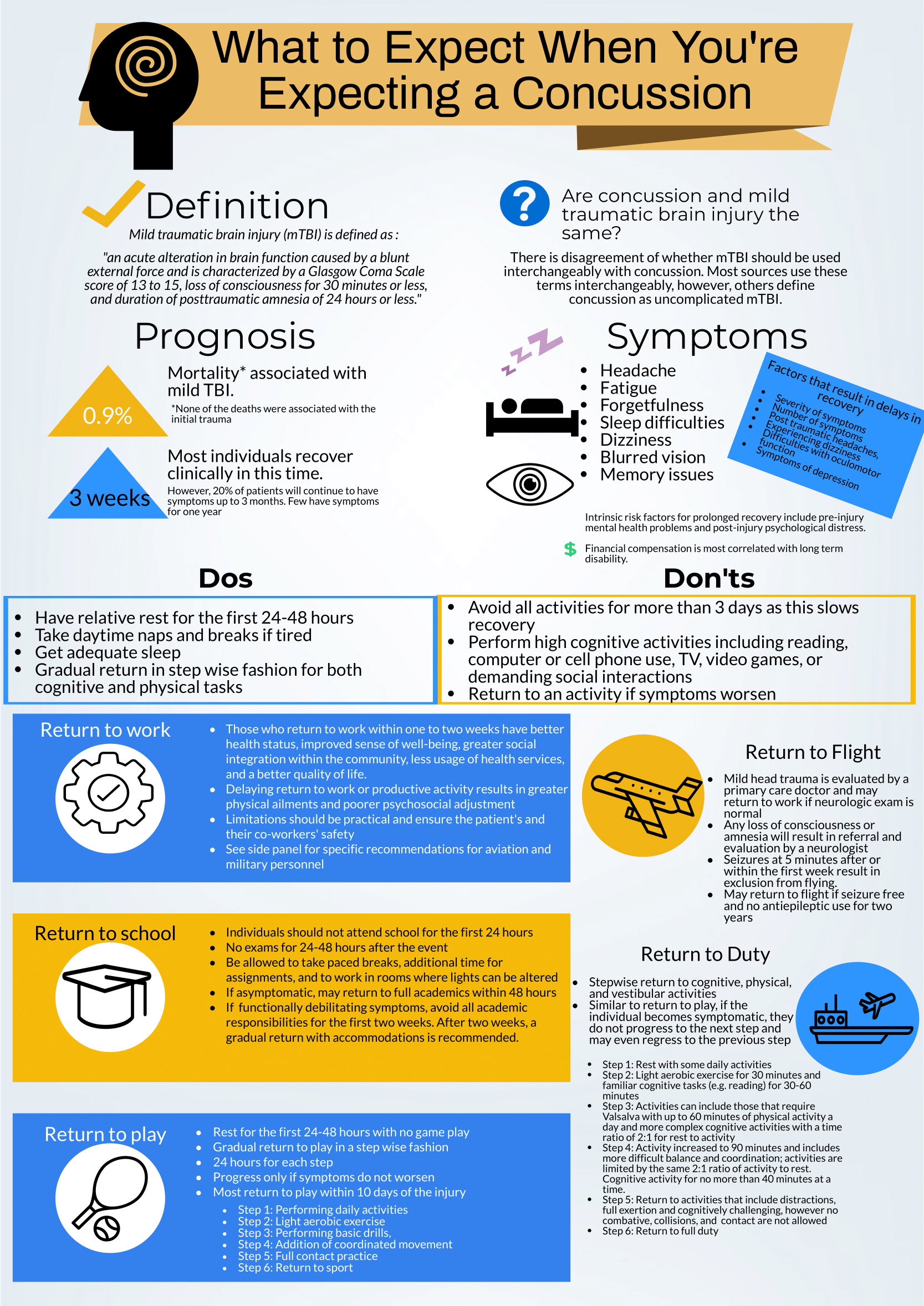
What to expect when you're expecting a concussion

Written by: Kelsey Green, MD (NUEM ‘23) Edited by: Jordan Maivelett, MD (NUEM ‘20) Expert Commentary by: Jake Stelter, MD

Written by: Kelsey Green, MD (NUEM ‘23) Edited by: Jordan Maivelett, MD (NUEM ‘20) Expert Commentary by: Jake Stelter, MD


Mild TBI Final Draft-1.png

Expert Commentary

This is a great review of anticipatory guidance when counseling patients who have been diagnosed with a concussion.  As noted, “mild traumatic brain injury (mTBI)” is often used synonymously with “concussion.” A better way to conceptualize this is to view concussion as a form of mTBI, realizing that mTBI can represent a spectrum of conditions.  One of the most important treatments of concussion from the Emergency Department (ED) perspective is to counsel patients on what to expect and how to best control their symptoms.  Concussions can present with a wide range of symptoms as detailed and can be quite distressing and disruptive to patients. As correctly pointed out, the presence of vestibular symptoms (i.e. dizziness or gait instability) as well as pre-existing mental health diagnoses, such as depression or anxiety, are associated with a protracted symptom course. Setting expectations of the symptoms they may develop and the possible timeline of symptom duration is important for patients as they manage their condition.  Early conservative treatment with adequate sleep and relative cognitive and physical rest will help manage and reduce the intensity of symptoms.  In our current society, it is nearly impossible to completely avoid screens and reading.  Hence, “everything in moderation” is appropriate when counseling these patients.  If the patient has to work at a computer, advise them to take frequent breaks for at least 10 minutes for every 30 minutes of screen time.  In addition, it is recommended that patients with a concussion avoid alcohol.  It is also advisable to avoid excessive caffeine.  However, if a patient already uses caffeine on a daily basis, they should not stop completely, as that can lead to withdrawal headaches.  Over-the-counter pain relievers, such as naproxen, ibuprofen or acetaminophen are appropriate for headache treatment, provided there are no contraindications to use.

 There are multiple return-to-learn, -work and -play protocols that have been published.  This is particularly applicable to athletes who have sustained a sport-related concussion (SRC).  Most schools and athletic programs have protocols that have been developed in conjunction with athletic trainers and team physicians.  It is important to remember that as an ED provider, you should not clear a patient to return to play.  That process needs to be conducted by the school athletic trainer in collaboration with the team physician after they have had the opportunity to evaluate the patient. You should consider referring your concussion patients to a Primary Care Sports Medicine or Neurology provider for follow-up if they do not have a team physician to visit.

There are multiple free resources available to providers who are interested in learning more about concussion and educating patients.  The Sport Concussion Assessment Tool – 5th Edition (SCAT5) is an in-depth evaluation tool that is often used by Sports Medicine clinicians when evaluating the extent and severity of a patient’s concussion syndrome.  These resources are listed here:

References

American Medical Society for Sports Medicine position statement on concussion in sport:

https://bjsm.bmj.com/content/53/4/213

SCAT5:

https://bjsm.bmj.com/content/bjsports/early/2017/04/26/bjsports-2017-097506SCAT5.full.pdf

27447 (1).jpg

Jacob Stelter, MD

Emergency Medicine, Primary Care Sports Medicine

Division of Emergency Medicine

NorthShore University HealthSystem


How To Cite This Post:

[Peer-Reviewed, Web Publication] Green, K. Maivelett, J. (2021, Feb 14). What to expect when you're expecting a concussion. [NUEM Blog. Expert Commentary by Stelter, J]. Retrieved from http://www.nuemblog.com/blog/concussion.


Posted on February 15, 2021 and filed under Neurology.

Marathon: The Collapsed Athlete

marathon image.png

Written by: Zach Schmitz, MD (PGY-3)  Edited by: Andrew Berg, MD (NUEM ‘19)  Expert Commentary by: Jake Stelter, MD


Marathon: The Collapsed Athlete

You’ve been enjoying a beautiful, 71 degree day wrapping ankles and rehydrating runners at the last marathon medical tent, just one mile from the finish. Suddenly, you get a different, more concerning type of call - there’s a runner down about a block south.

You fight against the flow of runners and finally see your patient on the left side of the course. He’s laying on his back and a bystander has placed an ice bag on his head. He tells you his name is Tony, but can’t tell you where he is or what he was doing. A friend says he was slowing down and looking unsteady before sitting on the curb. He’s sweating, working a little hard to breathe, and he has a 2+ radial pulse. What is your approach?

What to rule out first:

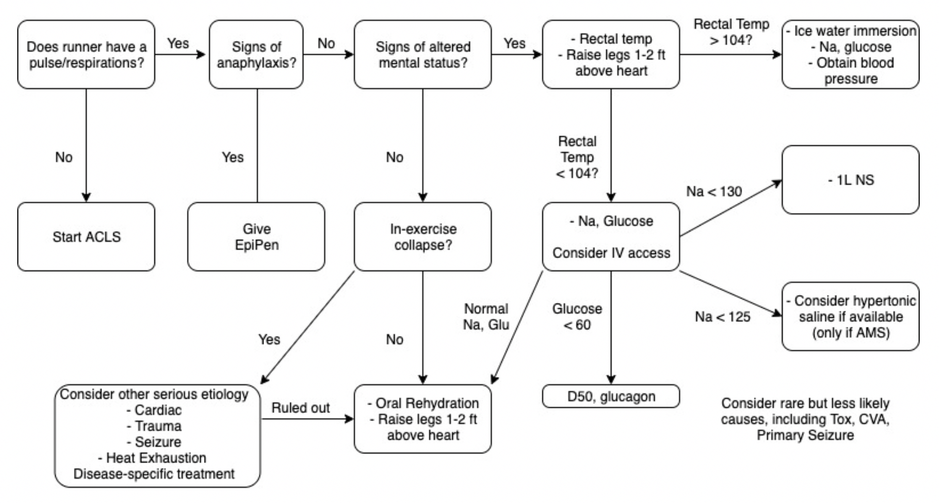
Just as with other ED patients, the first thing to do is rule out or intervene on life-threatening causes of runner’s collapse. Collapse during exercise is particularly concerning. There are five main causes of downed runners in that category: Sudden Cardiac Arrest, Exertional Heat Stroke, Anaphylaxis, Hypoglycemia, and Hyponatremia [1]. Below is an approach aimed toward addressing these concerns.

Marathon Algorithm.png

1. Sudden Cardiac Arrest

  • Suggested by an absent pulse and/or abnormal respirations.

  • Do not delay treatment. Start ACLS/BLS as your training and equipment allows and transport to a nearby ED.

  • Extremely rare[2]

2. Anaphylaxis

  • Suggested by any combination wheezes/stridor, shortness of breath, swelling, skin changes, nausea/vomiting, and altered mental status.

  • Treatment will likely be limited to IM epinephrine, as antihistamines, H2 blockers, and steroids are not routinely stocked in medical tents.

3. Exertional Heat Stroke

  • Suggested by altered mental status and a rectal temperature of > 104 degrees F.

  • These patients should be placed in an ice bucket immediately – any delay will risk permanent neurologic dysfunction.

  • Ice bucket immersion has been shown to reduce core body temperature 3x faster than ice towels and 15x faster than ice packs over major arteries[3].

  • Rapid on-site cooling is associated with better outcomes than immediate transfer to an emergency department for cooling. Those on site cooling end-points are controversial, but getting below 102 degrees F consistently leads to a safe transfer.[4]

  • You have to use rectal temperatures, as other temperature measurements have proven unreliable in a marathon setting.[4}

4. Hypoglycemia

  • Suggested by a spectrum from tremor, anxiety, diaphoresis, and altered mental status, up to seizure and coma.

  • Patients should be treated with glucose and transferred to a nearby medical facility.

5. Hyponatremia

  • Suggested by paresthesias, nausea/vomiting, and altered mental status, up to seizure and coma.

  • In one study of the Boston Marathon, 13% of runners had sodium values < 130, and 0.6% had critical values < 120. Those with longer race times, weight gain during the race, and those at the extreme ends of the BMI scale were more likely to have problems.[5]

  • Normal saline should be started for patients with initial Na of 130 or below, and 3% NS may be considered if Na < 125.


Thankfully, the above conditions comprise the minority of visits to medical tents at the marathon (including for downed runners). So what do you do with someone who is down and lightheaded but with a temperature of 99.3, sodium of 138, glucose of 98, and no signs of anaphylaxis? Collapse during exercise is still concerning, even after ruling out the causes above. You’ll want to confirm the patient can tolerate oral rehydration, place in a Trendelenberg position, and likely refer for further testing.

Collapse after exercise is more common, and, fortunately, often benign. Exercise associated collapse is likely to be the most frequent condition you encounter if you are in the final medical tent.


Exercise Associated Collapse (EAC)

Although considered to be more a chief complaint than diagnosis, EAC is defined as “a collapse in conscious athletes who are unable to stand or walk unaided as a result of light headedness, faintness and dizziness or syncope causing a collapse that occurs after completion of an exertional event.”[4] In one study, it accounted for 59% of patient presentations at the final medical tent.[1]

While running, increased oxygen demand by muscle leads to increased cardiac output and decreased peripheral vascular resistance. Skeletal muscle works as “second heart” for the race, overcoming this decrease in PVR to increase venous return. This mechanism is lost when running stops, and blood pools in the lower extremities. Cardiac output cannot be maintained, and perfusion is decreased. Further, the baroreceptor reflex controlling this mechanism is often compromised during long exercise. [6]

It is a fairly simple mechanism to reverse, and therefore a simple condition to treat. Placing the patient in a Trendelenberg position with the legs above the heart will usually achieve a fluid equilibrium in 10-30. Holtzhausen showed that these patients have no different electrolyte concentrations and are no more volume depleted than runners who finished the race without complication, so IV fluids are unnecessary[7]. However, keep in mind these people just ran a marathon, so they could probably use a little oral rehydration.

These patients should prove capable of sitting, then standing, then walking and eating before being discharged from the tent. If they show signs of altered mental status, vital sign abnormalities, or electrolyte imbalances, they should be treated appropriate and then transferred to an emergency department.


The vast majority of runners visiting a marathon medical tent are fully capable of finishing the race and just need help working out a cramp, covering up a blister, or grabbing some gel to cool a sore muscle. However, serious conditions do happen, and it is important to keep them in mind the next time you volunteer at your local marathon.


Take away points:

  • Life-threatening pathology is certainly possible in this relatively healthy cohort

  • Sudden cardiac arrest, exertional heat stroke, anaphylaxis, hypoglycemia, and hyponatremia should be considered for every down runner with altered mental status

  • Approach to the down runner: make sure you don’t need ACLS on scene > transfer to tent for rectal temp > Na, Glucose

  • If rectal temp is > 104, go directly to ice bath. Fully cool before transferring patient from tent

  • Although syncope post race can be scary, EAC is likely to resolve with 10-30 minutes of raised legs and oral rehydration


Expert Commentary

This is a great review of managing marathon runners who are acutely ill. It is important to keep in mind the diagnoses pointed out when dealing with a collapsed athlete.

1. Sudden Cardiac Arrest: This should be treated promptly following BLS/ACLS protocols. In this situation, the goal is to get to early defibrillation if possible as the most common cause is going to be a shockable arrhythmia, either ventricular fibrillation or ventricular tachycardia. The resources immediately available to you will vary depending on where on the course the patient goes down. Early activation of EMS is critical as they will bring with them both the means of transportation as well as ACLS supplies to aid in resuscitation.

2. Altered Mental Status: In a marathon athlete, the most important and life-threatening cause of altered mental status that needs to be ruled out is exertional heat stroke. As correctly pointed out, a core rectal temperature should be obtained on any athlete that is altered. Once identified as having a core temp over 103F, the athlete should be immediately cooled in an ice water tub until their temperature is 102F. At this point, the athlete should be removed from the water. Cooling below 102F can cause rebound hypothermia as cool peripheral blood shunts to the core. Avoid starting IV’s in runners prior to cooling, as getting blood into the tubs will contaminate them. If the athlete is normothermic and altered, check for hypoglycemia and treat accordingly.

3. Hyponatremia: Exercise-Induced Hyponatremia (EIN) is a relatively rare but very serious complication of endurance events. It is generally caused by excess sodium loss (sweating) that is often accompanied by excess free water intake. In a patient that is having signs and symptoms of neurologic dysfunction that is normothermic and not hypoglycemic, consider EIN. Common signs and symptoms include paresthesias, confusion, muscle weakness, cramping and seizures. If you have the ability to check a rapid sodium, then you can treat accordingly. If the patient’s sodium level is below 130 WITHOUT neurological symptoms, restrict free water intake and consider oral rehydration with electrolyte solutions. Sparingly administer isotonic IV fluids, no more than 250-500mL at a time and recheck the sodium level after each small bolus. If a patient falls into the category of hypervolemic hyponatremia, they may actually have an excess of ADH hormone and giving fluid may precipitate an even further drop in sodium. If a hyponatremic patient is having any neurological manifestation, especially seizures, the treatment is administration of 3% sodium chloride solution in 50-100mL boluses.

Also, as pointed out, be sure to consider other potential causes of your patient’s symptoms, including but not limited to cardiac pathology, trauma, stroke, and exercise associated collapse. Patients that are undifferentiated will often need to be transported to the nearest Emergency Department, as you are unlikely to have the resources to complete a diagnostic work-up in your course medical tent.

Jake Stelter.png

Jacob Stelter, MD

Instructor of Clinical Emergency Medicine

Primary Care Sports Medicine Fellow

University of Cincinnati

Medical Committee - Lead ICU Tent Coordinator

Bank of America Chicago Marathon


How to Cite This Post

[Peer-Reviewed, Web Publication] Schmitz Z, Berg, A. (2020, April 20). Marathon: The Collapsed Athlete. [NUEM Blog. Expert Commentary by Stelter, J]. Retrieved from http://www.nuemblog.com/blog/marathon


Other Posts You Might Enjoy:


References

[1] Roberts W, O’connor F, Grayzel J. Preparation and management of mass participation endurance sporting events. UpToDate. May 23 2017. https://www.uptodate.com/contents/preparation-and-management-of-mass-participation-endurance-sporting-events

[2] Roberts W.O., and Maron B.J.: Evidence for decreasing occurrence of sudden cardiac death associated with the marathon. J Am Coll Cardiol 2005; 46: pp. 1373-1374

[3] Casa D et al. Exertional Heat Stroke: New Concepts Regarding Cause and Care. Curr Sports Med Rep. 2012 May-Jun;11(3):115-23.

[4] Childress MA, O'Connor FG, Levine BD. Exertional collapse in the runner: evaluation and management in fieldside and office-based settings. Clin Sports Med. 2010 Jul;29(3):459-76. doi: 10.1016/j.csm.2010.03.007.

[5] Almond et al. Hyponatremia among runners in the Boston Marathon. N Engl J Med. 2005 Apr 14;352(15):1550-6.

[6] Asplund CA, O'Connor FG, Noakes TD Exercise-associated collapse: an evidence-based review and primer for clinicians Br J Sports Med 2011;45:1157-1162.

[7] Holtzhausen LM1, Noakes TD, Kroning B, de Klerk M, Roberts M, Emsley R. Clinical and biochemical characteristics of collapsed ultra-marathon runners. Med Sci Sports Exerc. 1994 Sep;26(9):1095-101.

[8] Madan S., Chung E., The Syncopal Athlete. American College of Cardiology. http://www.acc.org/latest-in-cardiology/articles/2016/04/29/19/06/the-syncopal-athlete. Apr 29 2016. Accessed 10 1 2017.

Posted on April 20, 2020 and filed under Environmental.